 한국학술지인용색인(NRF)


권호기사보기
| 기사명 | 저자명 | 페이지 | 원문 | 기사목차 |
|---|
| 대표형(전거형, Authority) | 생물정보 | 이형(異形, Variant) | 소속 | 직위 | 직업 | 활동분야 | 주기 | 서지 | |
|---|---|---|---|---|---|---|---|---|---|
| 연구/단체명을 입력해주세요. | |||||||||
|
|
|
|
|
|
* 주제를 선택하시면 검색 상세로 이동합니다.
배경: UIMD PBIA (ANI CO., 한국)는 새롭게 개발된 말초혈액 혈구 이미지의 자동화 분석장비이다. 본 연구에서는 UIMD PBIA의 백혈구 분류의 정확도와 처리속도를 평가하였다.
방법: 이상소견이 있는 환자 검체 192건과 이상소견이 없는 정상인 검체 50건을 포함한 총 242건의 말초혈액 도말로부터 얻어진 29,605개의 백혈구 세포 이미지를 이용하여 장비의 정확도와 처리 속도를 분석하였다.
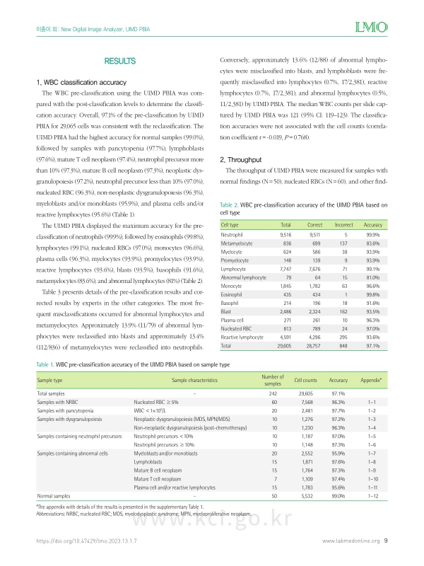
결과: UIMD PBIA는 정상인 검체에서 99%의 정확도를 보였고, 다섯 종류의 백혈구감별계산에서 99.2%의 정확도를 보였다. 오분류는 미성숙과립구, 모세포, 비정상 림프구에서 빈번하게 발생하여 이들에 대한 분석의 정확도는 81-93.9% 수준이었다. 비정상 혈구들은 같은 계열의 다른 세포로 분류되는 경향을 보였다. 장비의 처리속도는 시간당 42개 슬라이드였으며, 혈구감소증이 있는 경우는 시간당 29개 슬라이드였다.
결론: UIMD PBIA는 빠르고 정확한 백혈구 분류 결과를 제공하며, 특히 정상소견이나 혈구감소증이 있는 경우 유용하게 이용될 수 있을 것으로 생각된다.
Background: The UIMD PBIA (ANI CO., Suwon, Korea) is a newly developed automated digital image analyzer using innovative algorithms for the analysis of peripheral blood smears. We evaluated the accuracy and throughput of UIMD PBIA for the classification of white blood cells (WBCs).
Methods: A total of 29,605 cells in 242 clinical samples (192 samples with abnormal findings and 50 normal samples) were used to evaluate the classification accuracy and throughput of the UIMD PBIA. In addition, the total processing time for WBC classification by UIMD PBIA was measured to calculate the throughput.
Results: UIMD PBIA revealed outstanding performance for the identification of normal samples (99.0% accuracy) and five-part differentials (neutrophil, lymphocyte, monocyte, eosinophil, basophil, 99.2% accuracy). Misclassifications frequently occurred for immature granulocytes (83.6-93.9% accuracy), blasts (93.5% accuracy), and abnormal lymphocytes (81% accuracy). The pathogenic cells were likely to be misclassified into other classes of the same lineage. The average throughput was approximately 42 slides per hour. In cases with pancytopenia, the throughput was approximately 29 slides per hour.
Conclusions: The UIMD PBIA offers the most accurate results for WBC classification and the highest throughput, thereby reducing the technical workload, especially in cases with normal findings and pancytopenia. Accordingly, this study revealed the feasibility of using a digital switch in CBC analysis.| 번호 | 참고문헌 | 국회도서관 소장유무 |
|---|---|---|
| 1 | Clinical and Laboratory Standards Institute. Reference leukocytes (WBC) differential count (proportional) and evaluation of instrumental methods; Approved standard – Second edition. CLSI Document H20-A2. Wayne, PA: Clinical and Laboratory Standards Institute, 2007. | 미소장 |
| 2 | Da Costa L. Digital image analysis of blood cells. Clin Lab Med 2015;35:105-22. | 미소장 |
| 3 | Tatsumi N and Pierre RV. Automated image processing: past, present, and future of blood cell morphology identification. Clin Lab Med 2002;22:299-315. | 미소장 |
| 4 | Kratz A, Bengtsson HI, Casey JE, Keefe JM, Beatrice GH, Grzybek DY, et al. Performance evaluation of the CellaVision DM96 system: WBC differentials by automated digital image analysis supported by an artificial neural network. Am J Clin Pathol 2005;124:770-81. | 미소장 |
| 5 | Ceelie H, Dinkelaar RB, van Gelder W. Examination of peripheral blood films using automated microscopy; evaluation of Diffmaster Octavia and Cellavision DM96. J Clin Pathol 2007;60:72-9. | 미소장 |
| 6 | Briggs C, Longair I, Slavik M, Thwaite K, Mills R, Thavaraja V, et al. Can automated blood film analysis replace the manual differential? An evaluation of the CellaVision DM96 automated image analysis system. Int J Lab Hematol 2009;31:48-60. | 미소장 |
| 7 | Tabe Y, Yamamoto T, Maenou I, Nakai R, Idei M, Horii T, et al. Performance evaluation of the digital cell imaging analyzer DI-60 integrated into the fully automated Sysmex XN hematology analyzer system. Clin Chem Lab Med 2015;53:281-9. | 미소장 |
| 8 | Kim HN, Hur M, Kim H, Kim SW, Moon HW, Yun YM. Performance of automated digital cell imaging analyzer Sysmex DI-60. Clin Chem Lab Med 2017;56:94-102. | 미소장 |
| 9 | Kratz A, Lee SH, Zini G, Riedl JA, Hur M, Machin S, et al. Digital morphology analyzers in hematology: ICSH review and recommendations. Int J Lab Hematol 2019;41:437-47. | 미소장 |
| 10 | Fu X, Fu M, Li Q, Peng X, Lu J, Fang F, et al. Morphogo: an automatic bone marrow cell classification system on digital images analyzed by artificial intelligence. Acta Cytol 2020;64:588-96. | 미소장 |
| 11 | Palmer L, Briggs C, McFadden S, Zini G, Burthem J, Rozenberg G, et al. ICSH recommendations for the standardization of nomenclature and grading of peripheral blood cell morphological features. Int J Lab Hematol 2015;37:287-303. | 미소장 |
| 12 | Alférez E, Merino A, Mujica L, Rodellar J. Morphological features using digital image processing in lymphoid neoplasias. Int J Lab Hematol 2011;33(suppl 1):53. | 미소장 |
| 13 | Alférez S, Merino A, Bigorra L, Mujica L, Ruiz M, Rodellar J. Automatic recognition of atypical lymphoid cells from peripheral blood by digital image analysis. Am J Clin Pathol 2015;143:168-76. | 미소장 |
| 14 | Alférez S, Merino A, Mujica LE, Ruiz M, Bigorra L, Rodellar J. Automatic classification of atypical lymphoid B cells using digital blood image processing. Int J Lab Hematol 2014;36:472-80. | 미소장 |
| 15 | Bengtsson HI. Digital morphology analyzers in hematology: Comments on the ICSH review and recommendations. Int J Lab Hematol 2020;42:e213-5. | 미소장 |
| 16 | Kim AH, Lee W, Kim M, Kim Y, Han K. White blood cell differential counts in severely leukopenic samples: a comparative analysis of different solutions available in modern laboratory hematology. Blood Res 2014;49:120-6. | 미소장 |
*표시는 필수 입력사항입니다.
| *전화번호 | ※ '-' 없이 휴대폰번호를 입력하세요 |
|---|
| 기사명 | 저자명 | 페이지 | 원문 | 기사목차 |
|---|
| 번호 | 발행일자 | 권호명 | 제본정보 | 자료실 | 원문 | 신청 페이지 |
|---|
도서위치안내: / 서가번호:

우편복사 목록담기를 완료하였습니다.
*표시는 필수 입력사항입니다.
저장 되었습니다.
