Merkel cell carcinoma of the trunk : two case reports and imaging review = 몸통에 생긴 메르켈 세포암종 : 2예 증례 보고 및 영상 소견 고찰 / Ha Yun Oh ; Donghan Kim ; Yun Sun Choi ; EunKyung Kim ; Tae Eun Kim 1
[요약] 1
INTRODUCTION 1
CASE REPORT 2
CASE 1 2
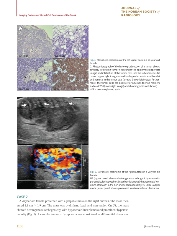
CASE 2 3
DISCUSSION 4
Author Contributions 5
REFERENCES 5
[요약] 6
초록보기
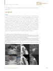
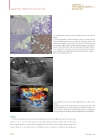
Merkel cell carcinoma (MCC) is a rare malignant cutaneous tumor primarily located in the head and neck. We report the imaging features of pathologically confirmed MCC in the trunk. On US, MCC showed heterogeneous echogenicity with perpendicular hypoechoic linear bands that resembled “columns of smoke” in the skin and subcutaneous layers as well as prominent vascularity. On MRI, the tumor showed hypointensity on T1-weighted images and hyperintensity on proton density and T2-weighted images with linear low-signal bands in the skin and subcutaneous layers as well as intense enhancement on T1-enhanced images. Although MCC has nonspecific imaging features, these characteristics may be helpful for the early diagnosis of this disease.
메르켈 세포암종은 드문 악성 피부종양으로 주로 두경부에 발생한다. 저자들은 몸통에 발생한 병리학적으로 확진된 메르켈 세포암종의 그레이스케일 및 색 도플러 초음파 소견과 자기공명영상 소견을 보고하고자 한다. 메르켈 세포암종은 초음파상 비균질 에코를 보이고 피부및 피하층에 수직방향으로 저에코의 ‘연기기둥’ 같은 선형 띠를 가지고 있었으며 내부에 혈류가 약간 증가되어 보였다. 한편 T1강조 자기공명영상에서 저신호강도, T2 및 양자밀도강조영상에서 같은 층에 저신호강도의 선형띠와 함께 고신호강도로 보였으며, 강한 조영증강을보였다. 메르켈 세포암종의 영상 소견이 비특이적으로 알려져 있지만, 이러한 특징적인 소견들은 이 질환을 조기 진단하는데 도움이 될 수 있을 것이다
 한국학술지인용색인(NRF)