
 대표어
대표어
 http://www.ndsl.kr/ndsl/search/detail/report/reportSearchResultDetail.do?cn=TRKO201700009458
권호기사보기
| 기사명 | 저자명 | 페이지 | 원문 | 기사목차 |
|---|
결과 내 검색
동의어 포함
표제지
목차
보고서 요약서 3
국문 요약문 4
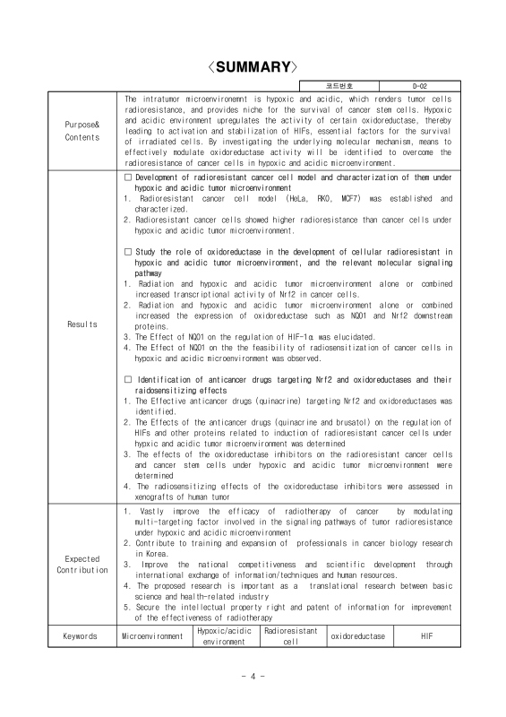
SUMMARY 5
제1장 연구개발과제의 개요 8
제1절 연구개발의 목적 8
제2절 연구개발의 필요성 8
1. 연구개발의 필요성 8
2. 연구개발 대상 기술의 경제적ㆍ산업적 중요성 및 연구개발의 필요성 9
제3절 연구개발의 범위 9
1. 연구목표 9
2. 연구개발의 범위 10
3. 연구개발의 개요 10
4. 연구개발의 추진체계 11
제2장 국내외 기술개발 현황 12
제1절 세계적 수준 12
제2절 국내 수준 12
제3절 국내외 연구현황 12
제4절 국내외 선행연구의 내용 및 결과 12
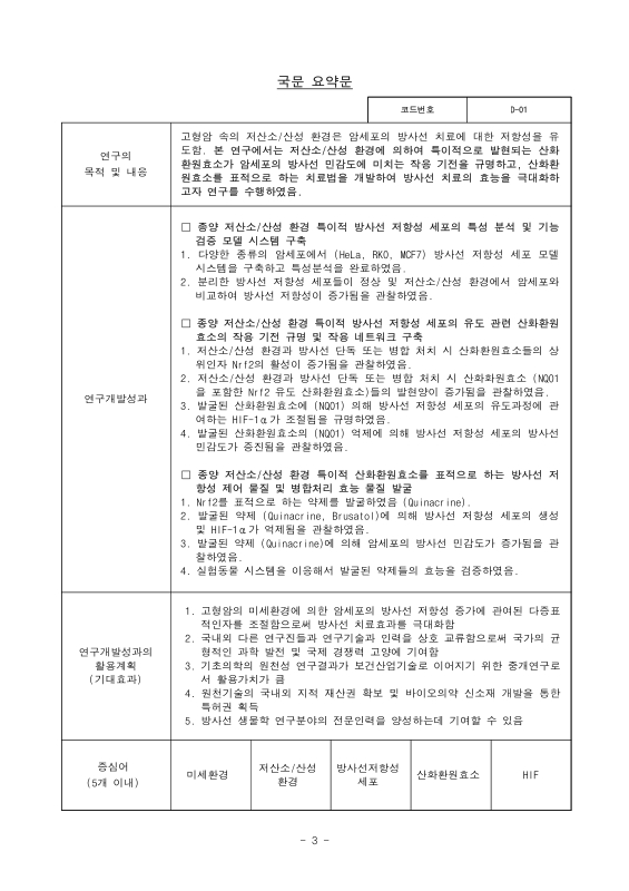
1. 고형암 암세포와 암줄기세포의 방사선 저항성과 관련된 미세환경 연구의 최근 동향 12
2. 방사선 치료에 미치는 저산소 및 산성 환경의 영향 13
3. 저산소 환경이 방사선 치료에 미치는 영향 13
4. 저산소 환경이 암세포의 방사선 저항성 증가에 미치는 영향 14
5. 산화적 스트레스 (oxidative stress) 15
6. 암줄기세포내의 산화환원효소의 발현 17
7. 암줄기 세포의 방사선 저항성 17
제5절 연구결과가 국내외 기술개발 현황에서 차지하는 위치 19
제3장 연구수행 내용 및 결과 20
1차년도 20
제1절 종양 저산소/산성 환경 특이적 방사선 저항성 세포의 특성 분석 및 기능 검증 모델 시스템 구축 20
제2절 종양 저산소 환경 특이적 방사선 저항성 세포의 유도 관련 산화환원효소의 작용 기전 규명 및 작용 네트워크 구축 22
2차년도 36
제1절 종양 저산소/산성 환경 특이적 방사선 저항성 세포의 특성 분석 및 기능 검증 모델 시스템 구축 36
제2절 종양 저산소 환경 특이적 방사선 저항성 세포의 유도 관련 산화환원효소의 작용 기전 규명 및 작용 네트워크 구축 37
3차년도 48
제1절 종양 저산소/산성 환경 특이적 산화환원효소를 표적으로 하는 방사선 저항성 제어 물질 및 병합처리 효능 물질 발굴 48
연구방법 56
제4장 목표달성도 및 관련분야 기여도 59
제1절 목표달성도 59
제2절 관련분야 기여도 60
제5장 연구결과의 활용계획 61
제1절 연구성과의 활용계획 61
제2절 타연구에의 응용 61
제6장 연구과정에서 수집한 해외과학기술정보 62
제1절 암 예방에 있어서 산화환원효소의 영향 보고 62
제2절 NQO1을 표적으로 하는 암치료 연구 보고 62
제7장 연구개발결과의 보안등급 63
제8장 국가과학기술종합정보시스템에 등록한 연구시설ㆍ장비 현황 64
제9장 연구개발과제 수행에 따른 연구실 등의 안전조치 이행실적 65
제1절 연구실 안전 점검 체계 및 실시 65
1. 실험실 안전 점검 체계 65
2. 실험실 정밀안전진단 실시 65
제2절 교육훈련 65
1. 개요 65
2. 교육과정ㆍ대상ㆍ내용 65
제3절 보험 가입 현황 66
제4절 기타 교육 및 안전관리 상황 66
제10장 연구개발과제의 대표적 연구실적 67
제11장 기타사항 68
제12장 참고문헌 69
그림 1. ISI Web of Knowledge, Web of Science search 13
그림 2. 암속의 저산소 빛 산성 환경 (Modified from Eric Hall, 1988) 13
그림 3. Survival curves for hypoxic and aerobic cells in cell culture (Rockwell et al., 2009) 13
그림 4. Radiation induced DNA damage under normoxia and hypoxia (Brown and Wilson, 2004) 14
그림 5. Schematic representation of various activators and inhibitors of reactive oxygen species production (Reuter et al., 2010) 14
그림 6. Cellular defense mechanisms against oxidative stress and hypoxia (Miyata et al., 2011) 15
그림 7. The signaling pathway induced by oxidative stresses (Dayem et al., 2010) 15
그림 8. Schematic representation of the Nrf2-Keap 1 defense pathway in renal tubular cells (Saito, 2013) 16
그림 9. The multiple cytoprotective functions of NQO1(Albena et a!. 2010) 17
그림 10. Signaling pathways that regulate self-renewal mechanisms during normal stem cell development and during transformation(Reya et al., 2011) 18
그림 11. Mechanisms of cancer stem cell radioresistance(Moncharmont et al., 2012) 18
그림 12. Construction of pEGFP-oDC1 20
그림 13. Isolation of cancer stem cell 20
그림 14. Isolation of radioresistance cells 20
그림 15. Characterization of cancer stem cell markers in cancer stem cell models 21
그림 16. Analysis of cancer stem cell markers using flow cytometry in MCF7, RKO and HeLa cancer stem cells 21
그림 17. Analysis of cancer stem cell markers using confocal microscopy in MCF7, RKO and HeLa cancer stem cells 21
그림 18. Clogenic surviving fraction of the cells with radiation under normoxic or hypoxia condition 22
그림 19. Clonogenic surviving fraction of the cells with radiation in RKO wild type and non-cancer stem cells under normoxic or hypoxia condition 22
그림 20. Translocation of Nrf2 in RKO cells treated with or without ionizing radiation (4 Gy) in neutral or acidic environment under normoxia or hypoxia 23
그림 21. Translocation of Nrf2 in RKO cells treated with or without ionizing radiation (4 Gy) in neutral or acidic environment under normoxia or hypoxia 23
그림 22. Analysis of Nrf2 activity in RKO cells treated with or without ionizing radiation (4 Gy) in neutral or acidic environment under normoxia or hypoxia 24
그림 23. Effect of hypoxia and ionizing radiation (4 Gy) on expression of NQO1 and Nrf2 target genes 25
그림 24. Effect of hypoxia and ionizing radiation (4 Gy) on expression of NQO1 and Nrf2 target proteins 25
그림 25. Effect of NQO1 on expression of HIFs 26
그림 26. Effect of NQO1 on expression of HIFs in various cancer cell lines 26
그림 27. Effect of NQO1 on expression of HIF-1 a 26
그림 28. Effect of NQO1 on expression of HIF-1 a regulatory proteins 27
그림 29. Effect of NQO1 on HIF-1 a stability under hypoxia 27
그림 30. Effect of NQO1 on ubiqutination of HIF-1 a under hypoxia 28
그림 31. Effect of NQO1 on proteasomal degradation of HIF-1 a under hypoxia 28
그림 32. Interaction between NQO1 and HIF-1 a under hypoxia 28
그림 33. NQOl interacts ODD domain of HlF-1 a under hypoxia 29
그림 34. Localization of NQO1 and HIF-1 a under normoxia and hypoxia 29
그림 35. Interaction between NQO1 and HIF-1 a under hypoxia 30
그림 36. Effect of NQO1 on interaction between HIF-1 a and E3 ligases 30
그림 37. Effect of NQO1 on Cullin5 mediated degradation of HIF-1 a under hypoxia 31
그림 38. Competition between NQO1 and Cullin5 for binding to HIF-1 a under hypoxia 31
그림 39. Effect of NQO1 on transactivation of HIF-1 a 32
그림 40. Effect of NQO1 on HIF-1 a induced VEGF expression under normoxia and hypoxia 32
그림 41. Effect of NQO1 on secretion of VEGF in cancer cells under normoxia and hypoxia 32
그림 42. A proposed model illustrating the effect of NQO1 on HIF-1 a 32
그림 43. Effect of NQO1 on expression of cancer stem cell markers in cancer cells 33
그림 44. Effect of NQO1 on generation of cancer stem cells in RKO cells treated with or without ionizing radiation (2 Gy) and hypoxia 33
그림 45. Effect of NQO1 on generation of cancer stem cells in various cancer cells treated with or without ionizing radiation (2 Gy) and hypoxia 34
그림 46. Effect of NQO1 on change of growth rates in cancer cells treated with or without ionizing radiation under normoxia and hypoxia 34
그림 47. Effect of NQO1 on clonogenic cell death in irracliated cancer cells 35
그림 48. Effect of oxidoreductases on generation of cancer stem cells in RKO cell under hypoxia 36
그림 49. Effect of oxidoreductases on expression of cancer stem cell marker (CD133) in RKO cell under hypoxia 36
그림 50. Effect of hypoxia and ionizing radiation (4 Gy) on expression of NQO1 and Nrf2 target genes 37
그림 51. Effect of hypoxia. acidosis and ionizing radiation (4 Gy) on expression of NQO1 and Nrf2 target protein. HO-1 37
그림 52. Effect of NQO1 on expression of HIFs 38
그림 53. Effect of NQO1 on expression of HIF-1 a 39
그림 54. Effect of NQO1 on expression of HIF-1 a regulatory proteins 39
그림 55. Effect of NQO1 on HIF-1 a stability under normoxia and hypoxia 40
그림 56. Interaction between NQO1 and HIF-1 a under hypoxia 40
그림 57. Localization of NQO1 and HIF-1 a under normoxia and hypoxia 41
그림 58. Effect of NQO1 on ubiqutination and proteasomal degradation of HIF-1 a 41
그림 59. Effect of NQO1 on interaction between HIF-1 a and PHD1-3 42
그림 60. NQO1 inhibits PHDs-mediated degradation of HIF-1 a in cancer cells under hypoxia 42
그림 61. Effect of NQO1 on transactivation of HIF-1 a 43
그림 62. Effect of NQO1 on tumor growth 44
그림 63. A proposed model illustrating the effect of NQO1 on HlF-1 a 44
그림 64. Effect of hypoxic and acidic microenvirorment on expression of HIF-1 a and cancer stem cell marker(CD133) 44
그림 65. Effect of NQO1 on change of growth rates in cancer cells treated with or without ionizing radiation under various oxygen concentration and pHs 45
그림 66. Effect of NQO1 on clonogenic cell death in irradiated cancer cells 45
그림 67. Effect of HO-1 on clonogenic cell death in irradiated cancer cells under normoxia and hypoxia 46
그림 68. Effect of Luteolin on expression of Nrf2 and HIF-1 a in cancer cells 46
그림 69. Luteolin-induced cancer cell death in normoxia and hypoxia 47
그림 70. Effect of anticancer drugs on transcripion activity of Nrf2 in cancer cells under hypoxia 48
그림 71. Effect of Nrf2 inhibitors on expression of NQO1 and Nrf2 target gene, HO-1 under hypoxia condition 49
그림 72. Effect of Nrf2 inhibitors on expression of HIF-1 a, Nrf2, NQO1 and Nrf2 target protein, HO-1 under hypoxia 49
그림 73. Effect of Nrf2 inhibitors on expression of HIF-1 a, Nrf2, NQO1 and Nrf2 target protein, HO-1 under hypoxia condition 49
그림 74. Effect of quinacrine on expression of HIF-1 a in colorectal cancer cells under hypoxia 50
그림 75. Effect of quinacrine on expression of HIF-1 a in colorectal cancer cells under hypoxia and radiation 50
그림 76. Effect of quinacrine on transactivation of HIF-1 a 50
그림 77. Effect of brusatol on expression of Nrt2 51
그림 78. Effect of brusatol on expression of HIF and its transcriptional activity in cancer cells under hypoxia 51
그림 79. Effect of brusatol on HIF-1 a protein stability in cancer cells under hypoxia 52
그림 80. PHDs requires brusatol-mediated degradation of HIF-1 a in cancer cells under hypoxia 52
그림 81. Effect of brusatol on activation of PHD in cancer cells under hypoxia 53
그림 82. Effect of quinacrine on change of growth rates in cancer cells treated with or without ionizing radiation under normoxia, hypoxia and acidic condition 53
그림 83. Effect of quinacrine on clonogenic cell death in irradiated cancer cells under hypoxia condition 54
그림 84. Effect of quinacrine on clonogenic cell death in irradiated cancer cells under normoxia, hypoxia and acidic condition 54
그림 85. Effect of quinacrine on IR-induced cell death in colorectal cancer cell under normoxia, hypoxia and acidic condition 54
그림 86. Combination effect of quinacrine and radiation on tumor growth using mouse xenograft model 55
그림 87. Effect of brusatol on tumor growth 55
*표시는 필수 입력사항입니다.
| 전화번호 |
|---|
| 기사명 | 저자명 | 페이지 | 원문 | 기사목차 |
|---|
| 번호 | 발행일자 | 권호명 | 제본정보 | 자료실 | 원문 | 신청 페이지 |
|---|
도서위치안내: / 서가번호:

우편복사 목록담기를 완료하였습니다.
*표시는 필수 입력사항입니다.
저장 되었습니다.
