
 대표어
대표어
 학술연구정보서비스(KERIS)

권호기사보기
| 기사명 | 저자명 | 페이지 | 원문 | 기사목차 |
|---|
결과 내 검색
동의어 포함
Title Page
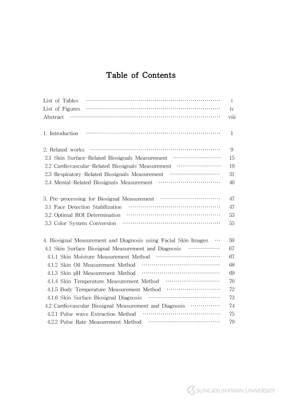
Contents
Abstract 14
1. Introduction 16
2. Related Works 24
2.1. Skin Surface-Related Biosignals Measurement 30
1) Contact devices based Skin surface-Related Biosignals Measurement 30
2) Non-Contact Skin surface-Related Biosignals Measurement 31
2.2. Cardiovascular-Related Biosignals Measurement 34
1) Contact devices based Cardiovascular-Related Biosignals Measurement 35
2) Non-Contact Cardiovascular-Related Biosignals Measurement 40
2.3. Respiratory-Related Biosignals Measurement 46
1) Contact devices based Respiratory-Related Biosignals Measurement 46
2) Non-Contact Respiratory-Related Biosignals Measurement 51
2.4. Mental-Related Biosignals Measurement 55
1) Contact devices based Mental-Related Biosignals Measurement 55
2) Non-Contact Mental-Related Biosignals Measurement 59
3. Pre-processing for Biosignal Measurement 62
3.1. Face Detection Stabilization 62
3.2. Optimal ROI Determination 68
3.3. Color System Conversion 70
4. Biosignal Measurement and Diagnosis using Facial Skin Images 74
4.1. Skin Surface Biosignal Measurement and Diagnosis 82
4.1.1. Skin Moisture Measurement Method 82
4.1.2. Skin Oil Measurement Method 83
4.1.3. Skin pH Measurement Method 84
4.1.4. Skin Temperature Measurement Method 85
4.1.5. Body Temperature Measurement Method 87
4.1.6. Skin Surface Biosignal Diagnosis 88
4.2. Cardiovascular Biosignal Measurement and Diagnosis 89
4.2.1. Pulse wave Extraction Method 90
4.2.2. Pulse Rate Measurement Method 94
4.2.3. Deep Learning based Pulse Rate Measurement Method 96
4.2.4. Blood Pressure Measurement Method 97
4.2.5. Paradoxical Pulse Measurement Method 106
4.2.6. Blood Volume Measurement Method 108
4.2.7. Hemoglobin Measurement Method 109
4.2.8. Blood Glucose Measurement Method 111
4.2.9. Blood Viscosity Measurement Method 113
4.2.10. Cardiovascular Biosignal Diagnosis 114
4.3. Respiratory Biosignal Measurement and Diagnosis 116
4.3.1. Respiratory signal Extraction Method 116
4.3.2. Respiration rate Measurement Method 119
4.3.3. Lung Function Measurement Method 121
4.3.4. Oxygen Saturation Measurement Method 126
4.3.5. Respiratory Biosignal Diagnosis 128
4.4. Mental Biosignal Measurement and Diagnosis 129
4.4.1. Stress Index Measurement Method 130
4.4.2. Depression disorder Index Measurement Method 136
4.4.3. Dementia Index Measurement Method 140
4.4.4. Mental Biosignal Diagnosis 141
5. Experiments and Implementations 142
5.1. Skin Surface Biosignal Measurement Experiment 143
5.1.1. Experimental results of Skin Moisture/Oil/pH Measurement 143
5.1.2. Experimental results of Skin Temperature Measurement 143
5.1.3. Experimental results of Body Temperature Measurement 144
5.2. Cardiovascular Biosignal Measurement Experiment 145
5.2.1. Experimental results of Pulse rate 145
5.2.2. Experimental results of Pulse rate using CNN & RNN 152
5.2.3. Experimental results of Blood pressure 153
5.2.4. Experimental results of Paradoxical pulse 158
5.2.5. Experimental results of Blood volume 158
5.2.6. Experimental results of Hemoglobin 159
5.2.7. Experimental results of Blood glucose 160
5.2.8. Experimental results of Blood viscosity 162
5.3. Respiratory Biosignal Measurement Experiment 163
5.3.1. Experimental results of Respiration rate 163
5.3.2. Experimental results of Lung function 168
5.3.3. Experimental results of Oxygen saturation 171
5.4. Mental Biosignal Measurement Experiment 172
5.4.1. Experimental results of Stress Index 172
5.4.2. Experimental results of Depression Index 178
5.4.3. Experimental results of Dementia 183
5.5. Integrated-Biosignal Measurement Application 185
6. Conclusions 188
References 193
논문요약 215
Fig. 1. Contact Devices-based Biosignal Measurement 25
Fig. 2. Different types of wearable devices 26
Fig. 3. Evolution of smart-phones over time 27
Fig. 4. Paradigm Transformation with the Development of Digital Video 28
Fig. 5. Biosignal Measurement Process Using Color Data of Facial and Parm skin Region of Interest (ROI) 29
Fig. 6. Examples of Wood's Lamp Usage 32
Fig. 7. Skin temperature measurement of the anterior body using Infrared Thermal Camera 33
Fig. 8. Block diagram of PPG device operation (a) A transimpedance amplifier stage that converts light intensity to an amplifier output voltage and (b) The... 36
Fig. 9. PPG signal containing Asystole event 38
Fig. 10. Pulse arrival time (PAT) and pulse transit time (PTT) 39
Fig. 11. Flowchart of the Pulse wave extraction and Pulse Rate measurement using the facial images based on ICA 41
Fig. 12. General contactless bio-signal measurement environment 42
Fig. 13. Participant posture and position of sensors to collect BP DB 44
Fig. 14. Overview of measurement of PTT and iPTT ((a) Contact method, (b) Non-contact method) 45
Fig. 15. Comparison of feature-based (red) and filter-based (green) techniques for extraction of exemplary respiratory signals during Inhalation and Exhalation 48
Fig. 16. Absorption spectra of hemoglobin 49
Fig. 17. Flowchart of the Respiratory signal extraction and Respiration Rate measurement using RGB color data of the facial images 52
Fig. 18. Flowchart of the Respiratory signal extraction and Respiration Rate measurement using RGB color data of the chest 52
Fig. 19. Illustration for traditional R and B channels method (AC components, where ACRed & ACblue are the standard deviations (SD) of the RGB data and...[이미지참조] 54
Fig. 20. Interval tachogram of 256 consecutive RR values in a normal subject at supine rest (a) and after head-up tilt (b). The HRV spectra are shown,... 57
Fig. 21. Typical Diastole series of PPG signal selected in each group(MDDSI+, MDDSI-, and CONT) 58
Fig. 22. Stress Classification using Facial Features, ECG, Voice 60
Fig. 23. Depression Classification using Pulse signal of facial images 61
Fig. 24. Expended set of Haar-like features: (a) edge features, (b) line features, (c) center-surround features. Face detection based on (d) the edge feature of... 65
Fig. 25. X-coordinate variability of ROI 66
Fig. 26. ROI stabilization process 66
Fig. 27. Calibrated x-coordinate variability of ROI (n=15) 67
Fig. 28. (a) Blood vessel and region of interest (red rectangle) of the facial artery. (b) Detected cheek ROI (green rectangle) from the facial video 69
Fig. 29. The extracted Cg signal recorded for 60s. 73
Fig. 30. Frequency Domain Distribution for BioSignal Measurement 76
Fig. 31. Simple block diagram of skin moisture/oil/ph analysis 82
Fig. 32. Simple block diagram of skin temperature 86
Fig. 33. Simple block diagram of Body temperature analysis 87
Fig. 34. Flowchart of Pulse wave Extraction 90
Fig. 35. Pulse-Frequency domain of the Cg signal (Fig. 29) 91
Fig. 36. Illustration of pulse wave extraction process 93
Fig. 37. Pulse wave comparison(PPG(blue), skin image(red)) 93
Fig. 38. Peak detection of pulse wave 94
Fig. 39. RR interval calculation of the pulse wave 95
Fig. 40. CNN based pulse rate classification pipeline 96
Fig. 41. RNN based pulse rate estimation pipeline 97
Fig. 42. Flowchart of systolic & diastolic blood pressure measurement 98
Fig. 43. Selection of two different ROIs for blood pressure measurement 99
Fig. 44. Peak detection and phase extraction of roi₁ and roi₂ 100
Fig. 45. Relationship dataset collection between the distance of camera-user and the detected face width 101
Fig. 46. Regression Analysis of Face width and Pixcel Distance 102
Fig. 47. Distance between roi₁ and roi₂ 103
Fig. 48. baPWV measurement 104
Fig. 49. PTT calculation using Pulse wave of roi₁ and roi₂ 105
Fig. 50. Pulse Wave Analysis in Inhalation and Exhalation Intervals 107
Fig. 51. Gradient calculation process 108
Fig. 52. Simple block diagram of Hemoglobin analysis 109
Fig. 53. Simple block diagram of Blood glucose analysis 111
Fig. 54. Blood viscosity calculation process 113
Fig. 55. Flowchart of Respiratory signal Extraction 117
Fig. 56. Respiration-Frequency domain of the Cg signal (Fig. 29) 118
Fig. 57. Illustration of respiratory signal extraction process 119
Fig. 58. Peak detection of respiratory signal 120
Fig. 59. PP interval calculation of the respiratory signal 120
Fig. 60. Simple block diagram of FVC, PEF, FVC(FEV₁, FEV₂ and FEV₃) analysis (Blow) 123
Fig. 61. Simple block diagram of FVC, PEF, FVC(FEV₁, FEV₂ and FEV₃) analysis (Non-blow) 125
Fig. 62. Flowchart of Activity Calculation 130
Fig. 63. A comparative analysis of changes in autonomic nervous system activity between healthy people (left) and depressed patient (right) 137
Fig. 64. Analysis of PCA application of frequency data for normal people and dementia patients (x-axis real part, y-axis imaginary part) 141
Fig. 65. Comparison of FPR calculation results by stable and unstable methods 146
Fig. 66. Graphs comparing FPR (Proposed) with other FPR methods (STFT, ACF, ICA-BPF) 148
Fig. 67. Comparison of BP calculation results by stable and original ROI 154
Fig. 68. Comparison of BP measurement results using PWV and PTT 156
Fig. 69. Comparison of FRR Results by face detection stabilization 163
Fig. 70. Comparison of FRR (Proposed) and other FRR (STFT, ACF, ICA-BPF) Graph 165
Fig. 71. Dementia index (x-axis) and correlation coefficient (y-axis) calculated for normal people and dementia patients 184
Fig. 72. Complexion state app (Measurement result screen) 185
Fig. 73. Lung function Measurement (Blow) 186
Fig. 74. Chart and management of Measured biosignal 187
본 논문에서는 얼굴 피부 영상을 이용한 비접촉 생체신호 측정 및 진단 방법을 제안한다. 이 논문은 네 가지 세부 연구 내용을 가지고 있다.
세부 연구 내용을 제안하기에 앞서, 조명 변화 및 신체 떨림 등으로 인해 발생하는 여러 노이즈를 제거하기 위해 얼굴 검출 안정화를 수행하고, RGB뿐만 아니라 YCgCo, YCbCr 등의 색차 성분을 이용한다. 또한, 제안된 방법을 통해 생체신호 측정 시스템의 성능이 향상된다는 것을 확인하였다.
첫째, 피부 관련 생체신호 측정 및 진단 방법을 제안한다. 제안된 접근법은 얼굴 영상의 피부 관심 영역에서 계산된 색상 및 질감 데이터를 이용하여 피부 수분, 피부 유분, 피부 산도 및 피부 온도를 측정하며, 얼굴 피부 영상을 이용하여 계산된 맥박수, 호흡수 및 피부 온도를 이용하여 맥박수, 호흡수 및 혈압을 포함하는 활력 징후의 필수 요소인 체온을 측정한다.
둘째는 심혈관 관련 생체신호 측정 및 진단 방법을 제안한다. 제안 방법은 다음과 같이 요약한다. (1) 혈당, 헤모글로빈은 피부 관심 영역에서 계산된 색상 데이터를 이용하여 측정한다. (2) 얼굴 영상의 피부 관심 영역에서 계산된 Cg 색상 신호로부터 산출된 맥파를 이용하여 맥박수, 기이맥, 부정맥 징후, 혈액점도, 혈량, 혈관 나이 및 혈과 탄성도를 측정한다.
셋째, 호흡기 관련 생체신호 측정 및 진단 방법을 제공하며, 얼굴 영상의 피부 관심 영역에서 계산된 Cg 색상 신호로부터 산출된 호흡 신호를 이용하여 호흡수, 폐기능(강제 호기량, 최대 호기량), 폐활량, 산소포화도를 측정한다.
넷째는 정신 건강 관련 생체신호 측정 및 진단 방법을 제안한다. 얼굴 영상에서 산출된 맥파와 호흡 신호를 분석하여 스트레스 및 우울증 지수를 측정하며, 전두엽 피부 관심 영역에서 계산된 Cg 색상 신호의 주파수 성분 분석을 통해 스트레스, 우울증 및 치매 지수를 측정한다. 제안된 주요 테마를 포함하는 얼굴 피부 영상을 이용한 비접촉 통합 생체신호 측정 및 진단 시스템 구축이 가능하다.*표시는 필수 입력사항입니다.
| 전화번호 |
|---|
| 기사명 | 저자명 | 페이지 | 원문 | 기사목차 |
|---|
| 번호 | 발행일자 | 권호명 | 제본정보 | 자료실 | 원문 | 신청 페이지 |
|---|
도서위치안내: / 서가번호:

우편복사 목록담기를 완료하였습니다.
*표시는 필수 입력사항입니다.
저장 되었습니다.
