
 대표어
대표어

권호기사보기
| 기사명 | 저자명 | 페이지 | 원문 | 기사목차 |
|---|
결과 내 검색
동의어 포함
표제지
요지
목차
제1장 서론 12
1.1. 연구 배경 및 목적 12
1.2. 연구 범위 15
제2장 갑상선 절제 후 치료준위 ¹³¹I을 투여한 암환자 주변의 방사선장 특성 18
2.1. 환자 주변 선량률 측정 18
2.2. 선량률 예측식 24
2.3. 불확도 27
제3장 방사성옥소 치료환자 퇴원 후 간병인의 피폭방사선량 평가 28
3.1. 방사성옥소 치료환자로 인한 제3자 피폭 이슈 28
3.2. 간병인 선량 측정방법 29
3.3. 간병인 선량 측정결과 31
3.4. 적정 표본의 수 35
제4장 방사성옥소 치료환자 퇴원 후 환자 간병인의 피폭방사선량 예측모델 38
4.1. 예측모델 유도 38
4.2. 간병인 선량에 영향을 미치는 인자 42
4.3. 환자 체내 방사성옥소의 기하학적 형태에 관한 고찰 43
4.4. 불확도 64
4.5. 간병 패턴 65
4.6. 입원 기간에 따른 비용_이득 분석 73
제5장 결론 및 건의 77
5.1. 결론 77
5.2. 고찰 및 건의 79
참고문헌 81
부록 8
부록 A. 방사성옥소-131 투여량 및 시간 추이에 따른 주위선량당량률 예측표 86
부록 B. 환자 입원기간 별 환자 가족 또는 간병인의 예상피폭선량(유효선량) 예측표 92
부록 C. 간병인 개인별 피폭선량 측정결과 및 도출된 간여인자 값 93
부록 D. 방사성옥소 ¹³¹I의 경구섭취에 따른 일반인의 전신 및 주요 장기별 섭취잔류분율 96
부록 E. 선량측정 기록지, 설문지 양식 및 선량계 사용요령서 100
ABSTRACT 105
Fig. 2.1. Measurement geometry of the external dose rates from patients administered radioiodine of therapy levels 19
Fig. 2.2. Survey meter (STEP, Germany, model OD-01HxE) used in the dose rate measurements. Equiped with an ion chamber suitable to measure 6 keV~3 MeV photon radiation 22
Fig. 2.3. Dose rates measured at 1 m in front of patients and their fit result as a function of time 26
Fig. 3.1. TLD (Theromo Luminescence Dosimeter) badge used in this study(Panasonic, Japan, model UD-874ATM). 32
Fig. 3.2. Frequency distribution of carer dose 34
Fig. 4.1. Pattern of ambient dose equivalent rate from patient 39
Fig. 4.2. Frequency distribution of engagement factor K. 46
Fig. 4.3. ICRP model for the bio-kinetics of radioiodine and removal constant 47
Fig. 4.4. Trend of intake retention fractions for ingestion of ¹³¹I as a function of time 48
Fig. 4.5. MIRD-ORNL phantom coronal image (created by MCNP program) 50
Fig. 4.6. ¹³¹I decay chart 52
Fig. 4.7. BOMAB phantom 56
Fig. 4.8. Image of the male computational phantom 57
Fig. 4.9. Irradiation geometry model from the BOMAB source phantom with uniform distribution to the target voxel phantom 59
Fig. 4.10. Irradiation geometry model from a point source(thyroid) in the BOMAB phantom to the target voxel phantom 60
Fig. 4.11. Comparison of dose rates to the carer from the point source model and the whole body distribution model 63
Fig. 4.12. Patterns of the deep dose Hp(10) to the carer of case A in every hours after the release of patient, measured by the active personal dosimeter 70
Fig. 4.13. Patterns of the deep dose Hp(10) to the carer of case B in every hours after the release of patient, measured by the active personal dosimeter 71
Fig. 4.14. Patterns of the deep dose Hp(10) to the carer of case C in every hours after the release of patient, measured by the active personal dosimeter 72
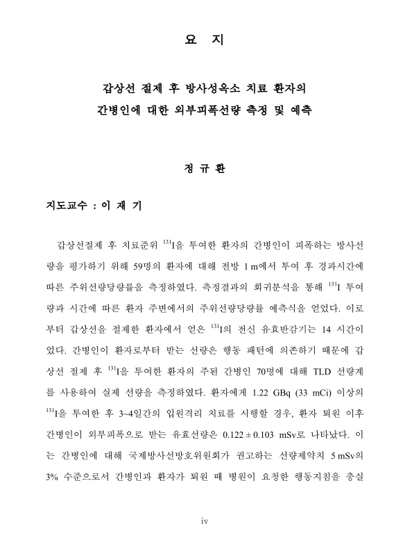
갑상선절제 후 치료준위 131I을 투여한 환자의 간병인이 피폭하는 방사선량을 평가하기 위해 59명의 환자에 대해 전방 1m에서 투여 후 경과시간에 따른 주위선량당량률을 측정하였다. 측정결과의 회귀분석을 통해 131I 투여량과 시간에 따른 환자 주변에서의 주위선량당량률 예측식을 얻었다. 이로부터 갑상선을 절제한 환자에서 얻은 131I의 전신 유효반감기는 14 시간이었다. 간병인이 환자로부터 받는 선량은 행동 패턴에 의존하기 때문에 갑상선 절제 후 131I을 투여한 환자의 주된 간병인 70명에 대해 TLD 선량계를 사용하여 실제 선량을 측정하였다. 환자에게 1.22 GBq (33 mCi) 이상의 131I을 투여한 후 3~4일간의 입원격리 치료를 시행할 경우, 환자 퇴원 이후 간병인이 외부피폭으로 받는 유효선량은 0.122±0.103 mSv로 나타났다. 이는 간병인에 대해 국제방사선방호위원회가 권고하는 선량제약치 5mSv의 3% 수준으로서 간병인과 환자가 퇴원 때 병원이 요청한 행동지침을 충실히 따른 것으로 평가된다. 이 결과로부터 131I Q0 GBq을 투여 받은 환자로부터 환자 퇴원 이후 전형적 간병인이 피폭할 근사적 개인선량은 0.86 KQ0e-0.05Tr mSv로 산출할 수 있음을 알았다. 여기서, 변수 Tr은 방사성옥소 투여 후 퇴원까지 시간(h)이며, K는 환자와 간병인이 접근하는 정도와 관련된 인자로서 1.29±0.88의 값을 가지는데 이를 '간여인자(engagement factor)'로 명명했다.
이 연구를 통해 개발된 모델에 근거하여 간병인이나 가족 구성원의 피폭 잠재성을 비용증가와 환자의 불편 증가와 대비함으로써 치료준위 131I을 투여한 갑상선 암환자의 격리입원 기간의 합리성을 재평가하였다. 그 결과 1.1GBq 이상 투여 환자에 대해 일률적으로 2박 3일 이상 입원 격리하는 현행 국내관행은 투여량이 7.4GBq 미만인 경우에는 과중한 것으로 나타나 투여 준위에 적합하게 조정하는 데 참조할 수 있는 기준표를 제안하였다. 퇴원 시점에서 환자 주변 주위선량당량률을 재확인하는 절차와 함께 이 기준표를 활용하면 국내 현안인 격리병실 부족문제를 완화하는 데 도움이 될 것이다. 제안된 절차는 간병인이나 성인 가족 구성원의 외부피폭에 근거하므로 이들의 유의한 내부피폭을 예방하는 지침이 필요하다. 민감 그룹인 아동의 피폭을 예방하는 서면지침도 요긴하다.| 번호 | 참고문헌 | 국회도서관 소장유무 |
|---|---|---|
| 1 | International Commission on Radiological Protection (1991). 1990 Recommendations of the International Commission of Radiological Protection. ICRP Publication 60. | 미소장 |
| 2 | International Atomic Energy Agency (2002). Radiological Protection for Medical Exposure to Ionizing Radiation, Safety Guide No. RS-G-1.5. IAEA, Vienna. | 미소장 |
| 3 | (2006). 국제방사선방호위원회 간행물 94: 비밀봉 방사성핵종으로 치료받은 환자의 퇴원, 한양대학교 방사선안전기술연구센터, 43-46. | 미소장 |
| 4 | U. S. Nuclear Regulatory Commission (1997). Code of Federal Regulations, 10 CFR Part 35.75, U. S. Government Printing Office, Washington DC. | 미소장 |
| 5 | U. S. Nuclear Regulatory Commission (1997). Release of Patients Administered Radioactive Materials, Regulatory Guide 8.39. | 미소장 |
| 6 | (2008). Consolidated Guidance About Materials Licenses - Program-Specific Guidance About Medical Use Licenses, Appendix U Model Procedure for Release of Patients or Human Research Subjects Administered Radioactive Materials, NUREG-1556, Vol.9, Rev. 2. | 미소장 |
| 7 | Radiation safety in the treatment of patients with thyroid diseases by radioiodine 131I : practice recommendations of the American Thyroid Association.  |
미소장 |
| 8 | Thyroid-Associated Paragangliomas |
미소장 |
| 9 | 원자력안전위원회 (2012.1.20). 고시 제2012-37호, 의료분야의 방사선 안 전관리에 관한 기술기준, 제12조 내지 제14조. | 미소장 |
| 10 | Radiation dose to family members of hyperthyroidism and thyroid cancer patients treated with 131I. |
미소장 |
| 11 | Could the treatment of differentiated thyroid carcinoma with 3.7 and 5.55 GBq of (131I)NaI, on an outpatient basis, be safe? |
미소장 |
| 12 | Relation between clinical and laboratory parameters with radiation dose rates from patients receiving iodine-131 therapy for thyroid carcinoma |
미소장 |
| 13 | Correlation between external exposure and activity in patients undergoing 131I thyroid cancer therapy. |
미소장 |
| 14 | (1995). Radiation Dose Rates from Patients Receiving Iodine-131 Therapy for Carcinoma of the Thyroid, EUR. J. Nucl. Med. 23:123-130. | 미소장 |
| 15 | 대한갑상선학회 (2012). 우리나라의 방사성 요오드 치료 현황과 안전관 리 -제언- 대한갑상선학회 춘계학술대회 발표자료, 2.24~2.25. | 미소장 |
| 16 | Effective Half-life of I-131 in Patients with Differentiated Thyroid Cancer Treated by Radioactive I-131 | 소장 |
| 17 | International Commission on Radiological Protection (1988). Individual Monitoring for Intakes of Radionuclides by Workers: Design and Interpretation. ICRP Publication 54, 141-154. | 미소장 |
| 18 | Measurements and prediction of the ambient dose rate from patient receiving radioiodine administration after thyroid ablation |
미소장 |
| 19 | Specific gamma-ray dose constants for nuclides important to dosimetry and radiological assessment |
미소장 |
| 20 | International Commission on Radiological Protection (1997). Individual Monitoring for Internal Exposure of Workers Replacement of ICRP Publication 54, ICRP Publication 78. | 미소장 |
| 21 | PREFACE |
미소장 |
| 22 | (2007). Int. Conf. on Nuclear Data for Science and Technology. | 미소장 |
| 23 | Health Physics Society (1995). Specifications for the Bottle Manikin Absorber Phantom. An American National Standard, ANSI/HPS N13.35, American National Standards Institute, New York. | 미소장 |
| 24 | Use of a voxel phantom as a source and a second voxel phantom as a target to calculate effective doses in individuals exposed to patients treated with 131I. |
미소장 |
| 25 | International Commission on Radiological Protection (2009). Adult Reference Computational Phantoms, ICRP Publication 110. | 미소장 |
| 26 | 4,6-Dimethyl-dibenzothiophene conversion over Al 2O 3–TiO 2-supported noble metal catalysts |
미소장 |
| 27 | (2011). 경수로형 원자력발전소 규제기준 및 규제지침, 규제기준, 제3장 설계공통 3.11.5 선량-금전계수, KINS/RS-N03.00, 77. | 미소장 |
| 28 | (1999). Internal Exposure to Relatives of Radioiodine Therapy Patients from I-131 Inhalation at Home, Radioactive Isotopes in Clinical Medicine and Research, XIII proceedings of the 23rd International Badgastein Symposium, 519-522. | 미소장 |
| 29 | 일본 후생성 통지 (1998.6.30). 방사성의약품이 투여된 환자의 퇴출(퇴원)에 대해서. | 미소장 |
| 30 | International Commission on Radiological Protection (1988). Radiation Dose to Patients from Radiopharmaceuticals. ICRP Publication 53, 275-278. | 미소장 |
*표시는 필수 입력사항입니다.
| 전화번호 |
|---|
| 기사명 | 저자명 | 페이지 | 원문 | 기사목차 |
|---|
| 번호 | 발행일자 | 권호명 | 제본정보 | 자료실 | 원문 | 신청 페이지 |
|---|
도서위치안내: / 서가번호:

우편복사 목록담기를 완료하였습니다.
*표시는 필수 입력사항입니다.
저장 되었습니다.
