
 대표어
대표어

권호기사보기
| 기사명 | 저자명 | 페이지 | 원문 | 기사목차 |
|---|
결과 내 검색
동의어 포함
표제지
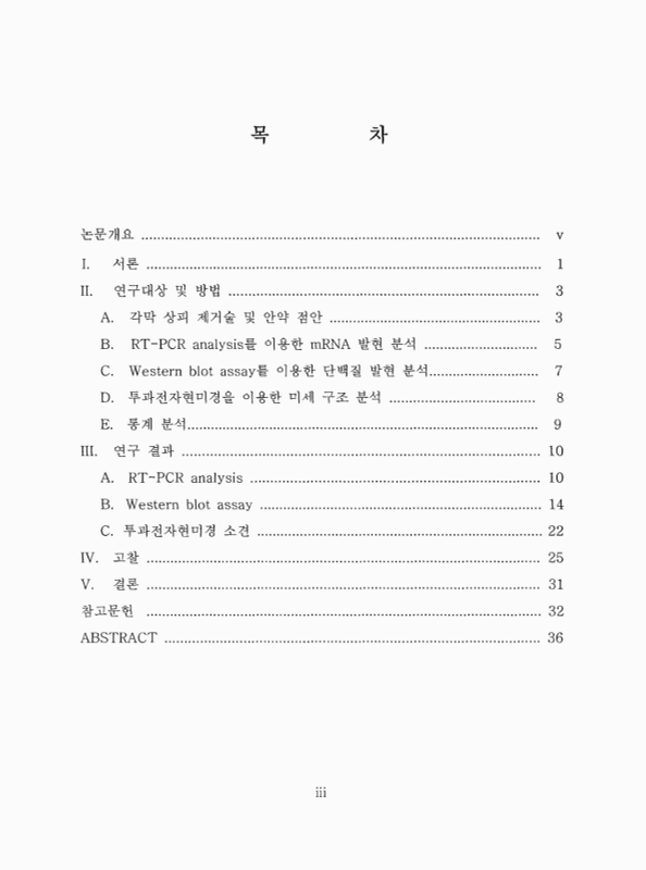
목차
논문개요 6
I. 서론 8
II. 연구 대상 및 방법 10
A. 각막 상피 제거술 및 안약 점안 10
B. RT-PCR analysis를 이용한 mRNA 발현 분석 12
C. Western blot assay를 이용한 단백질 발현 분석 14
D. 투과전자현미경을 이용한 미세 구조의 분석 15
E. 통계 분석 16
III. 연구 결과 17
A. RT-PCR analysis 17
B. Western blot analysis 21
C. 투과전자현미경 소견 29
IV. 고찰 32
V. 결론 38
참고문헌 39
ABSTRACT 43
Figure 1. RT-PCR of TGF-β₁ in rabbit corneas after treated with different fluoroquinolones. The expressions of TGF-β₁ were slightly higher in the debrided cornea but the differences were not significant (p>0.05,... 18
Figure 2. RT-PCR of IL-1α in rabbit corneas after treated with different fluoroquinolones. The expressions of IL-1α were higher in the debrided cornea than intact cornea in moxifloxacin, gatifloxacin and... 20
Figure 3. Western blot assay for MMP-1 in rabbit corneas after treated with different fluoroquinolones. Both in the debrided and intact cornea, the expressions of MMP-1 were upregulated in the gatifloxacin group more than... 22
Figure 4. Western blot assay for MMP-2 in rabbit corneas after treated with different fluoroquinolones. The expressions of MMP-2 were upregulated in debrided cornea than those in intact cornea in all test groups (p<0.05,... 24
Figure 5. Western blot assay for MMP-8 in rabbit corneas after treated with different fluoroquinolones. In the debrided cornea, expressions of the MMP-8 were upregulated in the intact cornea but the differences were not... 26
Figure 6. Western blot assay for MMP-9 in rabbit corneas after treated with different fluoroquinolones. The expressions of the MMP-9 were similar both in the debrided and intact cornea. Among the treated groups, there were... 28
Figure 7. Transmission electron micrographs of keratocytes treated with different fluoroquinolones. (A, B: Levofloxacin group; C, D: Moxifloxacin group; E, F: Gatifloxacin group) A. In the levofloxacin group,... 30
*표시는 필수 입력사항입니다.
| 전화번호 |
|---|
| 기사명 | 저자명 | 페이지 | 원문 | 기사목차 |
|---|
| 번호 | 발행일자 | 권호명 | 제본정보 | 자료실 | 원문 | 신청 페이지 |
|---|
도서위치안내: / 서가번호:

우편복사 목록담기를 완료하였습니다.
*표시는 필수 입력사항입니다.
저장 되었습니다.
