
 대표어
대표어
권호기사보기
| 기사명 | 저자명 | 페이지 | 원문 | 기사목차 |
|---|
결과 내 검색
동의어 포함
표제지
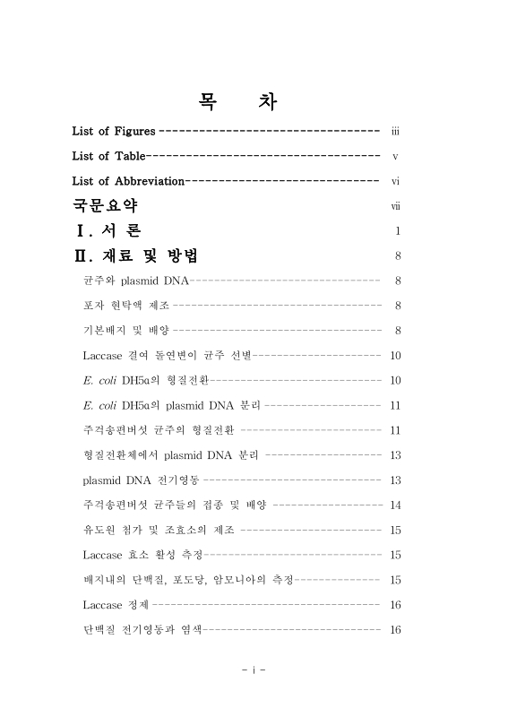
목차
List of Abbreviation 9
국문 요약 10
Ⅰ. 서론 11
Ⅱ. 재료 및 방법 18
균주와 plasmid DNA 18
포자 현탁액 제조 18
기본배지 및 배양 18
Laccase 결여 돌연변이 균주 선별 20
E. coli DH5α의 형질전환 20
E. coli DH5α의 Plasmid DNA 분리 21
주걱송편버섯 균주의 형질전환 21
형질전환체에서 plasmid DNA 분리 23
Plasmid DNA의 전기영동 23
주걱송편버섯 균주의 접종 및 배양 24
유도원 첨가 및 조효소의 제조 25
Laccase 효소 활성 측정 25
배지내 단백질, 포도당, 암모니아의 측정 25
Laccase 정제 26
전기영동과 염색 26
Ⅲ. 결과 27
Laccase 결여 돌연변이 균주 선정 27
주걱송편버섯 균주의 형질전환 28
Plasmid DNA 추출 및 확인 30
Laccase 생산에 미치는 유도원의 영향 31
배지내 단백질, 포도당, 암모니아의 변화 35
균주의 생장량 41
단백질 전기영동 43
Ⅳ. 고찰 45
Ⅴ. 참고문헌 49
ABSTRACT 58
Fig. 1a. Wild mushroom Pycnoporus cinnabarinus. 12
Fig. 1b. Growth of P. cinnabarinus on PDA medium. 12
Fig. 2. Structure of laccase active site. (adapted from Duran et al., 2002) 14
Fig. 3. Survival rate of the spores at U.V. irradiation; 30 W, 8cm distance from the U.V. bulb, mixing on a rotary shaker (50 rpm). 27
Fig. 4. Mutagenesis of P. cinnabarnus SCH-3 Wild type strain on PDA medium (0.02% sodium deoxycholate (pH 5.5) and 0.5 mM guaiacol). U.V. irradiation time is 120 s(A) and 150 s(B). M1 and M2 means laccase-deficient mutant. 28
Fig. 5. Growth of P. cinnabarinus strains on PDA medium 29
Fig. 6. Agarose gel electrophoresis of plasmid pELPL1 from E. coli DH5α and P. cinnabarinus strains. 30
Fig. 7. Time course of the laccase activity in the extracellular fluid of Pc-W. 31
Fig. 8. Time course of the laccase activity in the extracellular fluid of Pc-WP. 32
Fig. 9. Time course of the laccase activity in the extracellular fluid of Pc-MP. 32
Fig. 10a. Time course of the laccase activity in the extracellura fluid of P. cinnabarinus strains in the presence of 0.1 mM 2,5-xylidine as laccase inducer. 34
Fig. 10b. Time course of the laccase activity in the extracellura fluid of P. cinnabarinus strains in the presence of 1 mM ferulic acid as laccase inducer. 34
Fig. 10c. Time course of the laccase activity in the extracellura fluid of P. cinnabarinus strains in the presence of 25 g/L ethanol as laccase inducer. 35
Fig. 11. Time course of the protein productions in the extracellular fluid of Pc-W. 36
Fig. 12. Time course of the protein productions in the extracellular fluid of Pc-WP. 36
Fig. 13. Time course of the protein productions in the extracellular fluid of Pc-MP. 37
Fig. 14. Time course of the glucose concentrations in the extracellular fluid of Pc-W. 38
Fig. 15. Time course of the glucose concentrations in the extracellular fluid of Pc-WP. 38
Fig. 16. Time course of the glucose concentrations in the extracellular fluid of Pc-MP. 39
Fig. 17. Time course of the ammonia concentrations in the extracellural fluid of Pc-W. 40
Fig. 18. Time course of the ammonia concentrations in the extracellural fluid of Pc-WP. 40
Fig. 19. Time course of the ammonia concentrations in the extracellural fluid of Pc-MP. 41
Fig. 20a. Final biomass of P. cinnabarinus stains in the presence of 0.1 mM 2,5-xylidine 42
Fig. 20b. Final biomass of P. cinnabarinus stains in the presence of 1 mM ferulic acid 42
Fig. 20c. Final biomass of P. cinnabarinus stains in the presence of 25 g/L ethanol 43
Fig. 21. SDS-PAGE of laccase from P. cinnabarinus strains. 44
Fig. 22. Specific acitvity of P. cinnabarinus strains. 47
*표시는 필수 입력사항입니다.
| 전화번호 |
|---|
| 기사명 | 저자명 | 페이지 | 원문 | 기사목차 |
|---|
| 번호 | 발행일자 | 권호명 | 제본정보 | 자료실 | 원문 | 신청 페이지 |
|---|
도서위치안내: / 서가번호:

우편복사 목록담기를 완료하였습니다.
*표시는 필수 입력사항입니다.
저장 되었습니다.
