
 대표어
대표어

권호기사보기
| 기사명 | 저자명 | 페이지 | 원문 | 기사목차 |
|---|
결과 내 검색
동의어 포함
표제지
목차
I. 서론 11
II. 재료 및 방법 15
1. 실험재료 15
1) 시료 15
2) 시료 추출 및 분획 15
3) 시약 및 기기 18
2. 실험 방법 21
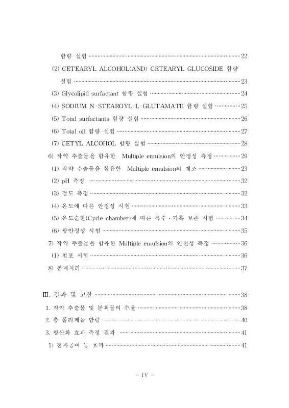
1) 총 폴리페놀 함량 측정 21
2) 항산화 효과 측정 22
3) 주름억제 효과 측정 24
4) 항균 효과 측정 29
5) Multiple emulsion 제조 및 실험 30
6) 작약 추출물을 함유한 Multiple emulsion의 안정성 측정 39
7) 작약 추출물을 함유한 Multiple emulsion의 안전성 측정 46
8) 통계처리 47
III. 결과 및 고찰 48
1. 작약 추출물 및 분획물의 수율 48
2. 총 폴리페놀 함량 50
3. 항산화 효과 측정 결과 51
1) 전자공여능 효과 51
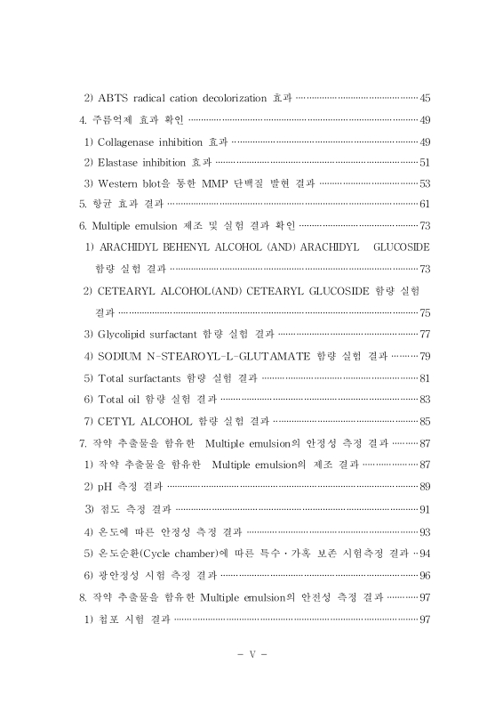
2) ABTS radical cation decolorization 효과 55
4. 주름억제 효과 확인 59
1) Collagenase inhibition 효과 59
2) Elastase inhibition 효과 61
3) Western blot을 통한 MMP 단백질 발현 효과 63
5. 항균 효과 결과 71
6. Multiple emulsion 제조 및 실험결과 확인 83
1) ARACHIDYL BEHENYL ALCOHOL (AND) ARACHIDYL GLUCOSIDE 함량실험 결과 83
2) CETEARYL ALCOHOL (AND) CETEARYL GLUCOSIDE 함량 실험 결과 85
3) Glycolipid surfactant 함량 실험 결과 87
4) SODIUM N-STEAROYL-L-GLUTAMATE 함량 실험결과 89
5) Total surfactants 함량 실험 결과 91
6) Total oil 함량 실험 결과 93
7) CETYL ALCOHOL 함량 실험 결과 95
7. 작약 추출물을 함유한 Multiple emulsion 안정성 측정 결과 97
1) 작약 추출물을 함유한 Multiple emulsion 제조 결과 97
2) pH 측정 결과 99
3) 점도 측정 결과 101
4) 온도에 따른 안정성 측정 결과 103
5) 온도순환(Cycle chamber)에 따른 특수·가혹 보존 시험측정 결과 104
6) 광 안정성 시험 측정 결과 106
8. 작약 추출물을 함유한 Multiple emulsion 안전성 측정 결과 107
1) 첩포 시험 결과 107
IV. 결론 109
V. 참고문헌 113
초록 119
Abstract 120
Fig. 1. The procedure for extraction from Paeonia lactiflora. 16
Fig. 2. The procedure for fraction from Paeonia lactiflora extracts. 17
Fig. 3. Preparation process of multiple emulsion in this experiments 31
Fig. 4. Preparation process of multiple emulsion containing Paeonia lactiflora extracts in this experiments. 40
Fig. 5. Cycle temperature change with day for 1 cycle. 44
Fig. 6. Electron donating ability of Electron donating ability of PLE. 53
Fig. 7. Electron donating ability of fractions from PLE. 54
Fig. 8. ABTS Free radical scavenging activity of PLE. 57
Fig. 9. ABTS Free radical scavenging activity of fractions from PLE. 58
Fig. 10. Collagenase inhibition activity of PLE. 60
Fig. 11. Elastase inhibition activity of PLE. 62
Fig. 12. Cell viability of PLE on HaCaT cell. 65
Fig. 13. Cell viability of fractions from PLE on HaCaT cell. 66
Fig. 14. Cell viability of PLE on Hs68 cell. 67
Fig. 15. MMP protein expression condition by UV-B on Hs68 cell. 69
Fig. 16. MMP13 protein expression inhibition by UV-B on Hs68 cell. 70
Fig. 17. The antimicrobial activity of PLE on microbial by disc diffusion method, respectively. 73
Fig. 18. The antimicrobial activity of PLE-D on microbial by disc diffusion method, respectively. 75
Fig. 19. The antimicrobial activity of PLE-E on microbial by disc diffusion method, respectively. 77
Fig. 20. The antimicrobial activity of PLE-B on microbial by disc diffusion method, respectively. 79
Fig. 21. The antimicrobial activity of PLE-W on microbial by disc diffusion method, respectively. 81
Fig. 22. Optical microscope images of multiple emulsion formulation A~D(x800) 84
Fig. 23. Optical microscope images of multiple emulsion formulation E~H(x800) 86
Fig. 24. Optical microscope images of multiple emulsion formulation I~L(x800) 88
Fig. 25. Optical microscope images of multiple emulsion formulation M~R(x800) 90
Fig. 26. Optical microscope images of multiple emulsion formulation S~Y(x800) 92
Fig. 27. Optical microscope images of multiple emulsion formulation AA~AE(x800) 94
Fig. 28. Optical microscope images of multiple emulsion formulation AF~AH(x800) 96
Fig. 29. Optical microscope images of multiple emulsion formulation AI(x800) 98
Fig. 30. Measurement of pH on the AI in the constant-temperature incubator. 100
Fig. 31. Measurement of viscosity on the AI in the constant-temperature incubator. 102
Fig. 32. Photograph of human patch test of AI. 108
Fig. 33. The mechanisms modulating the protective effect of Paeonia lactiflora extracts against UV-B 111
Fig. 34. The multiple emulsion diagram of surfactant and oil contents variation. 112
*표시는 필수 입력사항입니다.
| 전화번호 |
|---|
| 기사명 | 저자명 | 페이지 | 원문 | 기사목차 |
|---|
| 번호 | 발행일자 | 권호명 | 제본정보 | 자료실 | 원문 | 신청 페이지 |
|---|
도서위치안내: / 서가번호:

우편복사 목록담기를 완료하였습니다.
*표시는 필수 입력사항입니다.
저장 되었습니다.
