
권호기사보기
| 기사명 | 저자명 | 페이지 | 원문 | 기사목차 |
|---|
| 대표형(전거형, Authority) | 생물정보 | 이형(異形, Variant) | 소속 | 직위 | 직업 | 활동분야 | 주기 | 서지 | |
|---|---|---|---|---|---|---|---|---|---|
| 연구/단체명을 입력해주세요. | |||||||||
|
|
|
|
|
|
* 주제를 선택하시면 검색 상세로 이동합니다.
표제지
목차
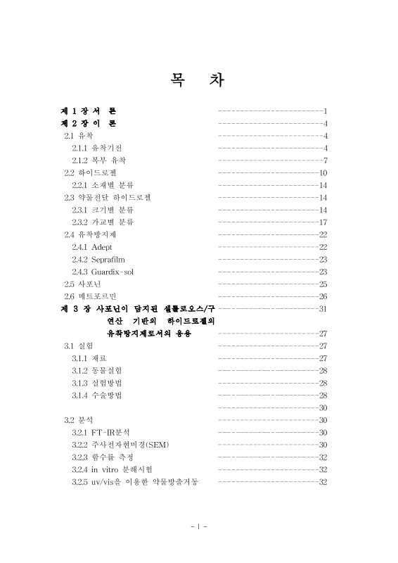
제1장 서론 10
제2장 이론 13
2.1. 유착 13
2.1.1. 유착기전 13
2.1.2. 복부 유착 16
2.2. 하이드로젤 19
2.2.1. 하이드로젤의 소재별 분류 21
2.3. 약물 전달 하이드로젤 23
2.3.1. 크기별 분류 23
2.3.2. 가교별 분류 26
2.4. 유착방지제 31
2.4.1. ADEPT 31
2.4.2. Seprafilm 32
2.4.3. Guardix-sol 32
2.5. 사포닌 34
2.6. 메트포르민 35
제3장 사포닌이 담지된 셀룰로오스/구연산 기반의 하이드로젤의 유착방지제로서의 응용 36
3.1. 실험 36
3.1.1. 재료 36
3.1.2. 동물실험 36
3.1.3. 실험방법 37
3.1.4. 수술방법 37
3.2. 분석 39
3.2.1. FT-IR 분석 39
3.2.2. 주사전자현미경 분석(SEM) 39
3.2.3. 함수율 측정 39
3.2.4. in vitro 분해 시험 40
3.2.5. UV/Vis를 이용한 약물 방출 거동 40
3.2.6. 유착 평가 40
3.2.7. 조직학적 절차 41
3.2.8. 면역조직화학염색법 41
3.2.9. 통계 분석 42
3.3. 결과 및 토론 43
3.3.1. CA-CS의 물리 화학적인 특징 43
3.3.2. CA-CS의 FTIR 분석 결과 45
3.3.3. CA-CS의 in vitro 생분해성 분석 결과 47
3.3.4. CA-CS의 SEM 촬영 분석 결과 49
3.3.5. CA-CS의 in-vitro 약물 방출 거동 분석 결과 51
3.3.6. CA-CS의 in-vivo 생분해성 분석 결과 53
3.3.7. 유착 조직의 면역화학염색법 분석 결과 55
3.3.8. 유착 조직의 면역형광염색법 분석 결과 57
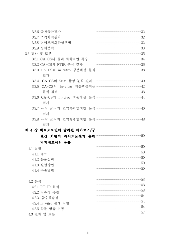
제4장 metformin이 담지된 agarose/citric ciacid 조성 유착방지제의 개발 59
4.1. 실험 59
4.1.1. 재료 59
4.1.2. 동물실험 59
4.1.3. 실험방법 60
4.1.4. 수술방법 60
4.2. 분석 62
4.2.1. FT-IR 분석 62
4.2.2. 접촉각 측정 62
4.2.3. 팽창 측정 62
4.2.4. in vitro 분해 시험 63
4.2.5. 약물방출거동 63
4.2.5. 유착 평가 63
4.2.6. 조직학적 절차 64
4.2.7. 면역조직화학염색 64
4.2.8. 통계 분석 64
4.3. 결과 및 토론 66
4.3.1. CA-AF의 물리 화학적인 특징 66
4.3.2. CA-AF의 FTIR 분석 69
4.3.3. CA-AF의 펭창율 결과 71
4.3.4. CA-AF의 표면접촉각 결과 73
4.3.5. CA-AF의 in-vitro 약물방출거동 겯과 75
4.3.6. 유착 육안 평가 결과 77
4.3.7. 유착 조직의 H&E염색 분석 결과 79
4.3.9. 유착 조직의 면역화학염색법 분석 결과 82
제5장 결론 84
5.1. 사포닌이 담지된 셀룰로오스/구연산 기반의 하이드로젤의 유착방지제로서의 응용 84
5.2. 메트포르민이 담지된 아가로스/구연산 기반의 하이드로젤의 유착방지제로서의 응용 85
참고문헌 87
ABSTRACT 90
Figure 1. Mechanism of normal healing and adhesion formation. 15
Figure 2. Schematic image of normal and adhesion. 17
Figure 3. Infection, surgery or trauma can cause adhesions to... 18
Figure 4. hydrogel of 3D-network structure. 20
Figure 5. delivery route of drug-eluting hydrogel 24
Figure 6. Mesh size mediates drug diffusion. 28
Figure 7. Chemical interactions mediate drug release 30
Figure 8. Preparation process of SAP@CA-CS 38
Figure 9. FT-IR spectra of CA-CS prepared with different weight of citric acid 46
Figure 10. in vitro degradation properties of CA-CS prepared with different... 48
Figure 11. SEM image of freeze-dried CA-CS with different concentrations of CA. 50
Figure 12. in vitro SAP-releasing properties of CA-CS cross-linked with... 52
Figure 13. In vivo biodegradability of SAP@CA-AF 54
Figure 14. Inhibition of formation of adhesion interface by Saponin@CA-CMC 56
Figure 15. Inhibition of fibroblast proliferation by Saponin@CA-CMC 58
Figure 16. Preparation process of MET@CA-AF 61
Figure 17. The novel synthesized MET@CA-CF. (A)The macroscopic... 67
Figure 18. FT-IR spectra of CA-AF prepared with different weight of citric acid 70
Figure 19. swelling ratio of CA-AF prepared with different weight of citric acid 72
Figure 20. contact angle of CA-AF prepared with... 74
Figure 21. in vitro MET-releasing properties of CA-AFs cross-linked with... 76
Figure 22. Attenuation of gross findings associated with postoperative... 80
Figure 23. Inhibition of the adhesion interface formation and myofibroblast activation by... 81
Figure 24. Inhibition of the adhesion interface formation and myofibroblast activation by... 83
*표시는 필수 입력사항입니다.
| 전화번호 |
|---|
| 기사명 | 저자명 | 페이지 | 원문 | 기사목차 |
|---|
| 번호 | 발행일자 | 권호명 | 제본정보 | 자료실 | 원문 | 신청 페이지 |
|---|
도서위치안내: / 서가번호:

우편복사 목록담기를 완료하였습니다.
*표시는 필수 입력사항입니다.
저장 되었습니다.
