
 대표어
대표어
권호기사보기
| 기사명 | 저자명 | 페이지 | 원문 | 기사목차 |
|---|
결과 내 검색
동의어 포함
Title Page
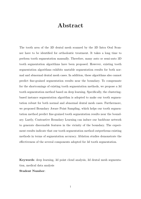
Abstract
Contents
Chapter 1. Introduction 9
1.1. Related work 11
1.1.1. Deep learning based 3d model analysis 11
1.1.2. 3D point cloud instance segmentation 12
1.1.3. 3D dental model segmentation 13
Chapter 2. Proposed Method 14
2.1. Overview 14
2.2. Tooth Group Network 15
2.3. Boundary Aware Point Sampling 21
2.4. Tooth type classification 22
2.5. Training details 22
Chapter 3. Experimental Results 24
3.1. Dataset 24
3.2. Evaluation metrics 24
3.3. Comparison with other methods 25
3.4. Ablation studies 29
3.5. robustness to abnormal case 32
Chapter 4. Discussion 36
Chapter 5. Conclusion 38
Bibliography 39
초록 44
Figure 2.1. (a) Our tooth segmentation pipeline predicts tooth instance labels for each vertex of the dental mesh. (b) Point Group Module takes sampled point cloud and outputs tooth type labels and tooth instance labels. These... 16
Figure 2.2. The entire architecture of the Point Transformer used in Point Group Network. Sampled point cloud(n x 6) is fed into the Point Transformer in the... 17
Figure 2.3. After DBSCAN[29], we calculate the variance of each cluster. If the variance of a certain cluster is more than 3 times higher than the average... 18
Figure 2.4. (a) The red area means incorrect tooth instance labels while the green area means correct tooth instance labels. (b) visualization of the concept... 19
Figure 2.5. The red points represent the sampled points obtained using the Farthest Point Sampling, while the green points correspond to the unsampled... 21
Figure 3.1. Qualitative comparison results. 27
Figure 3.2. Visualization of the tooth segmentation results with or without Boundary Aware Point Sampling. 31
Figure 3.3. Visualization of the tooth segmentation results with or without Tooth Group Network. 32
Figure 3.4. Visualization of the tooth segmentation results with or without Mask Refinement Module. 33
Figure 3.5. Ablation study for Boundary Aware Point Sampling. 34
Figure 3.6. Abnormal dental cases. (a) missing teeth. (b) misaligned teeth. (c) raw dental mesh without preprocessing. (d) Teeth equipped with braces. 35
치과에서 환자의 치아를 교정하거나 수복하는 치료에 대한 계획을 세우기 위해, 치기공사들은 CAD 툴을 이용하여 3차원 구강 스캐너를 통해 수집된 치아 매시 데이터에서 각 치아 영역을 분리해야 한다. 3차원 치아 매시 데이터에서 각 치아를 분리해내는 일은 시간이 많이 소요되는 작업이며, 이를 보완하기 위해 자동/반자동 치아 분할 알고리즘이 연구됐다. 하지만, 이전까지 제안된 알고리즘들은 상실된 치아가 있는 경우나 교정기가 부착되어있는 경우 등 특이 케이스에 대해 잘 동작하지 않는 경우가 있었으며, 평범한 구강 스캔 데이터라도 불안정한 분할 결과를 내놓는 경우가 많았다. 또한, 치아와 잇몸 사이의 경계나 치아와 치아 사이의 경계와 가까운 부분에서 잘못된 분할 결과를 내놓는 경우가 많았다.
본 논문에서는 기존 치아 분할 알고리즘의 단점을 보완하기 위한 인공지능 네트워크를 제안한다. 구체적으로, 클러스터링 방식의 인스턴스 분할 방법을 적용하여 상실 치아가 있는 경우나 교정기가 부착된 케이스 등 다양한 케이스에서 올바른 치아 분할 결과를 도출할 수 있도록 네트워크를 구성하였다. 그리고, 대조 경계 학습을 적용하여 치아와 치아사이의 경계와 치아와 잇몸 사이의 경계 부분에서 치아 분할성능을 높였다. 또한, 경계 인식 포인트 샘플링 기법을 제안하고, 이를 이용해 경계 부분에서 더 많은 매시의 정점을 샘플링함으로써 치아 분할 성능을 높였다.
본 논문에서 제안한 치아 분할 방법이 기존에 제안된 다른 방법보다 더 좋은 성능을 가짐을 실험적으로 확인하였다. 또한, 추가 실험을 통해 우리가 치아 분할 알고리즘에 도입한 각 요소들이 효과가 있음을 보였다.*표시는 필수 입력사항입니다.
| 전화번호 |
|---|
| 기사명 | 저자명 | 페이지 | 원문 | 기사목차 |
|---|
| 번호 | 발행일자 | 권호명 | 제본정보 | 자료실 | 원문 | 신청 페이지 |
|---|
도서위치안내: / 서가번호:

우편복사 목록담기를 완료하였습니다.
*표시는 필수 입력사항입니다.
저장 되었습니다.
