
 대표어
대표어
 학술연구정보서비스(KERIS)

권호기사보기
| 기사명 | 저자명 | 페이지 | 원문 | 기사목차 |
|---|
결과 내 검색
동의어 포함
Title Page
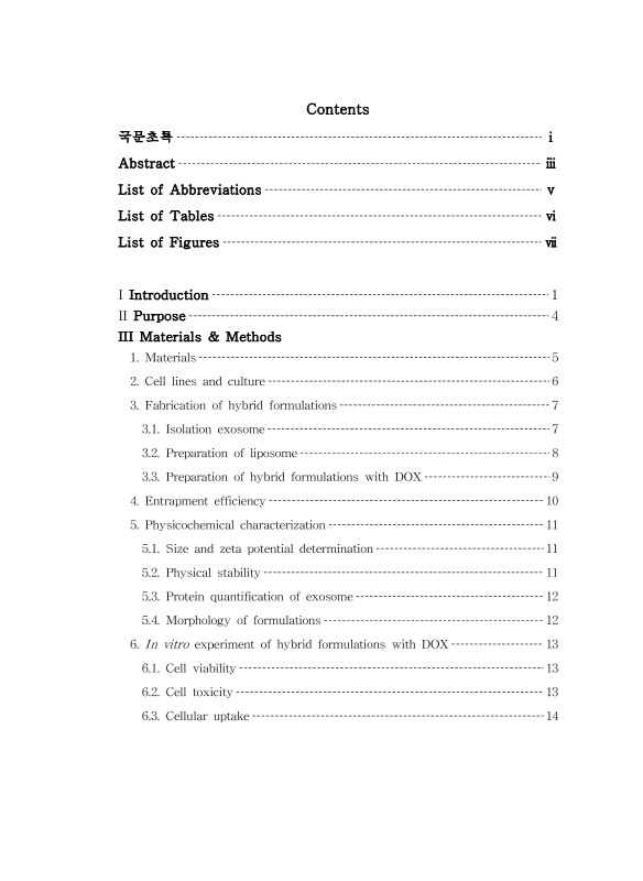
Contents
국문초록 7
ABSTRACT 9
List of Abbreviation 11
Ⅰ. Introduction 15
Ⅱ. Purpose 18
Ⅲ. Materials & Methods 19
1. Materials 19
2. Cell lines and culture 20
3. Fabrication of hybrid formulations 21
3.1. Isolation exosome 21
3.2. Preparation of liposome 22
3.3. Preparation of hybrid formulations with DOX 23
4. Entrapment Efficiency 24
5. Physicochemical characterization 25
5.1. Size and zeta potential determination 25
5.2. Physical stability 25
5.3. Protein quantification of exosome 26
5.4. Morphology of formulations 26
6. In vitro experiment of hybrid formulations with DOX 27
6.1. Cell viability 27
6.2. Cell toxicity 27
6.3. Cellular uptake 28
Ⅳ. Results & Discussion 29
1. Characterization of exosome 29
2. Characterization of hybrid formulation 31
2.1. Size, zeta potential and encapsulation efficiency 31
2.2. Physical stability 37
3. In vitro experiment of hybrid formulations with DOX 43
3.1. Cell viability 43
3.2. Cell toxicity 50
3.3. Cellular internalization 54
Ⅴ. Conclusions 58
Ⅵ. References 59
Figure 1A. Cryo-TEM images of hybrid formulations (a: F1, b: F2, c: F3, d: F4) 36
Figure 2A. The Percentage of relative size of hybrid formulations in a saline 39
Figure 2B. The Percentage of relative size of hybrid formulations in a 10% FBS (*p〈.05.) 40
Figure 2C. Physical stability of hybrid formulations in Triton X-100 41
Figure 2D. The Percentage of relative size of hybrid formulations in Triton X-100 42
Figure 3A. MCF-7 cell image (KCLB NO.30022) 44
Figure 3B. MDA-MB-231 cell image (ATCC® No.HTB-26™)[이미지참조] 45
Figure 3C. MDA-MB-468 cell image (ATCC® No.HTB-132™)[이미지참조] 46
Figure 4A. Cell viability of hybrid formulations without the drug in MCF-7 cells for 24 h 47
Figure 4B. Cell viability of hybrid formulations without the drug in MDA-MB-231 cells for 24 h 48
Figure 4C. Cell viability of hybrid formulations without the drug in MDA-MB-468 cells for 24 h 49
Figure 5A. Cell toxicity of hybrid formulations with the drug in MCF-7, MDA-MB-231 and MDA-MB-468 for 4 h at 37 ℃... 52
Figure 5B. Cell toxicity of hybrid formulations with drug in MCF-7 for 4 h at 4 ℃ and 37 ℃ (*p〈.05.) 53
Figure 6A. CLSM of hybrid formulations with drug in MCF-7 at 37 ℃ 55
Figure 6B. CLSM of hybrid formulations with drug in MDA-MB-231 at 37 ℃ 56
Figure 6C. CLSM of hybrid formulations with drug in MDA-MB-468 at 37 ℃ 57
*표시는 필수 입력사항입니다.
| 전화번호 |
|---|
| 기사명 | 저자명 | 페이지 | 원문 | 기사목차 |
|---|
| 번호 | 발행일자 | 권호명 | 제본정보 | 자료실 | 원문 | 신청 페이지 |
|---|
도서위치안내: / 서가번호:

우편복사 목록담기를 완료하였습니다.
*표시는 필수 입력사항입니다.
저장 되었습니다.
