
 대표어
대표어





권호기사보기
| 기사명 | 저자명 | 페이지 | 원문 | 기사목차 |
|---|
결과 내 검색
동의어 포함
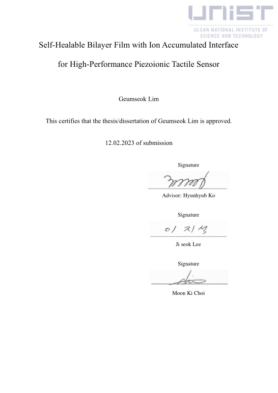
Title Page
Abstract
Contents
Ⅰ. Introduction 15
1.1. Iontronic skin 15
1.2. Ion migration mechanism in flexible iontronics 17
1.3. Multifunctional iontronic tactile sensors 18
1.4. Application of iontronic tactile sensors 21
1.5. Recent challenges of iontronic tactile sensors 23
1.6. Research goal 26
Ⅱ. Experimental Method 36
2.1. Preparation of cross-linked PVDF-HFP/ion liquid composite 36
2.2. Preparation of piezoionic tactile sensor array 37
2.3. Characterization 38
Ⅲ. Result and Discussion 42
3.1. Fabrication of bilayerd piezoionic tactile sensor 42
3.2. Piezoionic performance 46
3.3. Comparison of piezoionic performance between single-layer and bilayer devices 48
3.4. Characterization of piezoionic performance 51
3.5. Application of piezoionic tactile sensor arrays 53
Ⅳ. Conclusion 69
Ⅴ. References 70
Figure 1.1. (a) Overview of interfacial iontronic sensing (Y. Chang et al. Adv. Mater., 2021, 33, 2003464). Schematic illustrating the ion migration by (b) voltage (Y. Chang et al. Adv. Mater., 2021,... 28
Figure 1.2. (a) Ion transport mechanism called as "hopping" that ions jump to another site including intra- and inter-chain of polymer (Z. Xue et al. J. Mater. Chem. A. 2015, 3, 19218). (b) Schematic... 29
Figure 1.3. Various types of multifunctional iontronic tactile sensors. (a) Decoupled multimodal temperature, strain sensor (I. You et al. Science, 2020, 370, 961). (b) Ion-dipole interaction based... 30
Figure 1.4.1. e-IPC for artificial skin.. (a) Multicelluar hybrid ionic pump (V. Amoli et al. Nat. Commun. 2019, 10, 4019). Various types of ionic sensor arrays (C. Pang et al. Nat. Mater. 2012, 11, 795) (b)... 31
Figure 1.4.2. Human-interactive technologies equipment. a) Sensing glove (A. Atalay et al. Adv. Mater. Technol. 2017, 2, 1700136). b) Monitoring performance of human arms movement angle and speed (C.... 32
Figure 1.4.3. Wearable healthcare monitoring devices. (a) Human pulse signal monitoring (A. Atalay et al. Adv. Mater. Technol. 2017, 2, 1700136). (b) Human breath monitoring (C. Walsh et al. Adv.... 33
Figure 1.5. Recent advances for improvement of sensing performance of I-skin. (a) (N. Bai et al. Nat. Commun., 2020, 11, 209). (b) (E. Boahen et al. Nat. Commun., 2022, 13, 7699). (c) (P. Zhang et al.... 34
Figure 1.6. Schematics illustrating the comprehensive concepts of bilayer piezoionic tactile sensors. (a) The operating mechanisms for (i) ion-dipole interactive self-healing bilayer and (ii) ion accumulated... 35
Figure 2.1. Fabrication process of crosslinked ionic PVDF-HFP elastomer. 39
Figure 2.2. (a) Photograph of ionic PVDF-HFP elastomer with various ion liquid contents from 0 wt% to 20 wt%. Material analysis of (b) stress-strain curve, corresponding (c) stretchability, (d) Young's... 40
Figure 2.3. (a) Photograph of unstable interface of overlapped two films and (b) corresponding piezoionic output signals. (c) Schematic of the sample preparation for self-healing test. Sample is... 41
Figure 3.1. The corresponding (a) self-healing behavior and (b) piezoionic property recognizing bending direction. 54
Figure 3.2. (a) Synthesis process of crosslinked PVDF-HFP elastomer and (b) corresponding enhanced mechanical properties. Material analysis for (c) self-healing efficiency, (d) AC frequency-dependent... 54
Figure 3.3. FT-IR spectra of (a) neat and crosslinked PVDF-HFP elastomer, (b) EMIM-TFSI, PVDF-HFP and PVDF-HFP/EMIM-TFSI composite. 55
Figure 3.4. (a) Photograph of unstable interface of overlapped two films and (b) corresponding piezoionic output signals. (c) Schematic of the sample preparation for self-healing test. Sample is... 56
Figure 3.5. Fabrication process of piezoionic tactile sensor array (1 x 4 pixels and each sensor size showing 5 x 5 mm²). 57
Figure 3.6. EIS analysis of ionic composite with varying ion contents. (a) Nyquist plot and fitted curve including a semicircle and straight tail corresponding bulk film and EDL, respectively. (b) Schematic... 58
Figure 3.7. (a-c) Bode plots with various ion concentrations. Blue circle is overlapped point between real and imaginary impedance curve, that frequency indicates the charge relaxation frequency. 58
Figure 3.8. Piezoionic performance of bilayer devices. (a) Photographs showing the sensor responding to bending stimuli, (b) schematic illustrations of the ion distribution during bending, and (c)... 59
Figure 3.9. Illustration demonstrating the assessment of piezoionic characteristics and ion distribution across the electrodes in response to bending stimuli, leading to variances in electrode potential and the... 60
Figure 3.10. (a-c) Cyclic voltammetry plots generated by applying voltages from -1 V to 1 V with varying ion concentrations, and corresponding (d) a table summarizing current and power density at 1V. 60
Figure 3.11. A comparative analysis between single-layer and bilayer devices, featuring (a, d) cross-sectional film images post-cross-cutting, and electrical responses during bending stimuli, including (b,... 61
Figure 3.12. Comparison of piezoionic performance between single- and bilayer devices. (a) The current response of single-layer devices under bending deformation and (b) FFT wave pattern for line-... 62
Figure 3.13. EIS analysis for the comparison of ion movement in single- and bilayer films. Nyquist plots representing (a) the single-layer and (b) the bilayer film under different DC biases, along with (c)... 63
Figure 3.14. Cyclic voltammetry plots generated by applying voltages ranging from -1 V to 1 V for (a) bilayer and (b) single-layer film. (c) A table summarizing current and power density at 1V. 64
Figure 3.15. Comparative analysis of three distinct multi-layered devices with double (thickness of 710 μm), triple (740 μm), and quadruple (700 μm) stacking configurations. Cross-sectional optical images... 65
Figure 3.16. Perception of static and dynamic deformations for surface texture discrimination and braille reading. (a) Schematic illustrates the static perception of gradual bending deformation, and (b)... 66
Figure 3.17. (a-c) Optical images showing top view of 3D-printed line patterns, and corresponding FFT results after scanning various pitches of line pattern (1500, 1000, 500 µm). 67
Figure 3.18. (a) Photograph of 3D-printed braille with the word "ionic sensor" and (b) cross-sectional image representing embossed braille. (c) Optical images of braille surfaces with three different... 67
*표시는 필수 입력사항입니다.
| 전화번호 |
|---|
| 기사명 | 저자명 | 페이지 | 원문 | 기사목차 |
|---|
| 번호 | 발행일자 | 권호명 | 제본정보 | 자료실 | 원문 | 신청 페이지 |
|---|
도서위치안내: / 서가번호:

우편복사 목록담기를 완료하였습니다.
*표시는 필수 입력사항입니다.
저장 되었습니다.
