
 대표어
대표어
권호기사보기
| 기사명 | 저자명 | 페이지 | 원문 | 기사목차 |
|---|
결과 내 검색
동의어 포함
목차
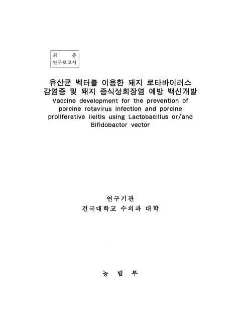
표제지=0,1,1
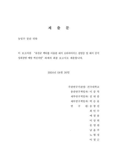
제출문=0,2,1
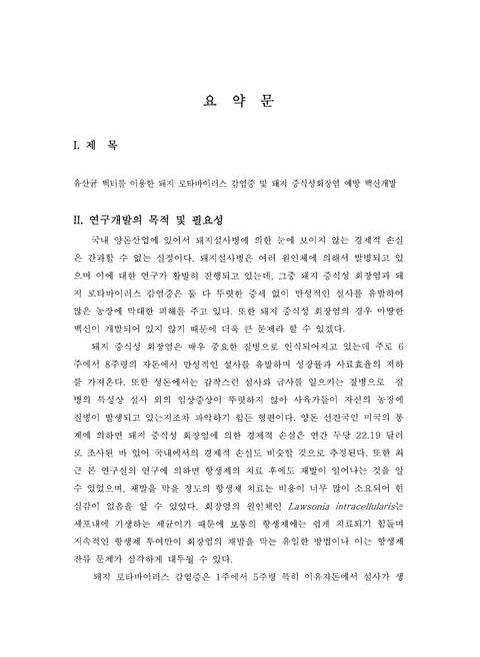
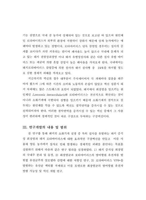
요약문=0,3,5
Summary=0,8,3
Contents=0,11,3
목차=0,14,3
제1장 연구 개발과제의 개요=1,17,4
제2장 국내외 기술개발 현황=5,21,1
제3장 연구개발수행 내용 및 결과=6,22,1
제1절 돼지증식성회장염균의 분리 및 동정=6,22,1
1. 서론=6,22,3
2. 재료 및 방법=9,25,1
가. 돼지증식성회장염균의 분리 및 배양=9,25,1
1) 분리재료=9,25,1
2) 조직배양=9,25,2
3) 장조직유제의 세포 감염=10,26,1
4) 감염 세포조직의 맹복적 계대배양=10,26,2
5) 감염 세포조직의 감염 여부 관찰=11,27,2
6) 면역조직화학염색=12,28,1
나. 돼지 증식성회장염의 재현성 실험=12,28,1
1) 실험동물=12,28,2
2) 조직배양된 돼지증식성회장균의 준비=13,29,1
3) 장조직유제추출균의 준비=13,29,2
4) 접종량의 결정=14,30,1
5) 접종=14,30,1
6) 임상증상의 평가기준설정=15,31,1
7) 분변에서의 균체검사=15,31,1
8) 혈청학적 검사=15,31,1
9) 간접형광항체법=16,32,1
10) 부검=16,32,1
11) 조직학적 검사=16,32,1
3. 결과=17,33,1
가. 국내에서 분리된 Lawsonia균의 분리 및 배양=17,33,1
(1) 균 분리에 사용된 재료=17,33,3
(2) Lawsonia균의 분리=19,35,1
(3) Lawsonia균의 유지배양=19,35,3
(4) 분리균주의 동정=21,37,2
나. 돼지 증식성 회장염의 재현=22,38,1
(1) 임상증상=22,38,2
(2) 분변내 Lawsonia 균 검출 및 혈중 항체가 검사=23,39,3
(3) 부검 소견=26,42,1
(4) 면역조직화학 염색 결과=26,42,3
다. 햄스터에서의 증식성 회장염 유도=28,44,3
라. 국내 돼지증식성회장염의 감염 분포상황=30,46,3
4. 고찰=33,49,2
제2절 유산균의 Salmonellar균 감염 방어능 비교=35,51,1
1. 서론=35,51,2
2. 재료 및 방법=37,53,1
가. 실험동물=37,53,1
나. 유산균 급여=37,53,1
다. 살모넬라 감염에 대한 유산균 급여 마우스의 방어효과=37,53,1
라. 비장세포 증식 시험=37,53,2
마. 탐식세포 활성화 시험=38,54,1
바. 장관내 분비형 IgA의 정량 분석=38,54,1
사. 면역화학적 검사=38,54,2
아. 통계학적 분석=39,55,1
3. 결과=40,56,1
가. 비피도박터 유산균의 살모넬라균 감염에 대한 방어효과=40,56,2
나. 비피도박터 유산균의 비장 T와 B세포 활성화 효과=41,57,2
다. 비피도박터 유산균의 탐식세포 활성화 효과=42,58,2
라. 비피도박터 유산균의 장내 분비형 IgA 중강 효과=44,60,2
4. 고찰=46,62,3
제3절 유산균을 이용한 돼지로타바이러스 VP8* 발현=49,65,1
1. 서론=49,65,3
2. 재료 및 방법=52,68,1
가. 세균의 종류 및 배양 배지=52,68,1
나. 플라스미드와 DNA 조작=52,68,2
다. 셔틀벡터 작제 pNZ123:SPO2/B-galactosidase=53,69,1
라. Lactobacillus와 Bifidobacterium에서 발현할 셔틀벡터에 VP8*의 크로닝=53,69,2
마. 전기영동과 Wester blot=54,70,2
3. 결과=56,72,1
가. Shuttle vector pNZ123:SPO2:beta-galactosidase의 제작=56,72,1
나. XL1-Blue 에서의 beta-galactosidase의 발현=56,72,1
다. Shuttle vector pBESAF2/VP8*의 발현=56,72,1
라. E. coli와 Bifidobacteria에서 VP8*의 발현=56,72,2
마. Shuttle vector pAT:pgsA/VP8*의 제작=57,73,1
바. E. coli와 Lactobacilus casei에서 VP8*의 발현=57,73,6
4. 고찰=63,79,2
제4장 목표달성도 및 관련분야에의 기여도=65,81,1
제5장 연구개발결과의 활용계획=66,82,2
제6장 연구개발과정에서 수집한 해외과학기술정보=68,84,1
제7장 참고문헌=69,85,16
영문목차
[title page etc.]=0,1,10
Contents=0,11,6
I. Introduction of project=1,17,4
II. General consideration of ralated technology=5,21,1
III. Results of the research=6,22,1
A. Isolation and identification of Lawsonia intracellularis=6,22,1
1. Introduction=6,22,3
2. Materials and methods=9,25,1
2.1. Isolation and in vitro cultivation of L. intracellularis=9,25,1
2.1.1. Source material=9,25,1
2.1.2. Cell culture=9,25,2
2.1.3. Infection of cell culture with intestine mucosal homogenate=10,26,1
2.1.4. Blind passage of the infected cells=10,26,2
2.1.5. Monitoring of infection=11,27,2
2.1.6. Immunohistochemistry=12,28,1
2.2. Experimental induction of PE=12,28,1
2.2.1. Animals=12,28,2
2.2.2. Preparation of L. intracellulars pure culture inoculum=13,29,1
2.2.3. Preparation of intestinal mucosa homogenate inoculum=13,29,2
2.2.4. Quantificantion of inoculum=14,30,1
2.2.5. Inoculation=14,30,1
2.2.6. Clinical observation=15,31,1
2.2.7. Fecal shedding tests(PCR)=15,31,1
2.2.8. Serology testing=15,31,1
2.2.9. Indirect fluorescent antibody tests(1FAT)=16,32,1
2.2.10. Necropsy and evaluation of macroscopic lesions=16,32,1
2.2.11. Histologic tests=16,32,1
3. Results=17,33,1
3.1. In vitro cultivation and maintenance of L. intracellularis=17,33,1
3.1.1. Source material used in isolation of L. Intracellularis=17,33,3
3.1.2. Isolation of L. intracellularis=19,35,1
3.1.3. Cultivation and maintenance=19,35,3
3.1.4. Confirmation of the isolate=21,37,2
3.2. Experiment 1-Experment reporduction of PE=22,38,1
3.2.1. Clinical results=22,38,2
3.2.2. PCR and serum IgG titer=23,39,3
3.2.3. Macroscopic lesions=26,42,1
3.2.4. Immunohistochemistry staining=26,42,3
3.3. Experiment 2-Induction of PE in hamsters=28,44,3
3.4. Prevalence of PPE in Korea=30,46,3
4. Discussion=33,49,2
B. Comparison of prtective potentials between bifidobacterium longum and Lactobacillus casei against Salmonella Infection=35,51,1
1. Introduction=35,51,2
2. Materials and methods=37,53,1
2.1.1. Animals=37,53,1
2.1.2. Feeding procedure=37,53,1
2.1.3. Protective effect test against Salmonella infection in mice=37,53,1
2.1.4. Spleen cellproliferation assay=37,53,2
2.1.5. Macrophage activation assay=38,54,1
2.1.6. Determination of the level of sIgA from the intestinal contents=38,54,1
2.1.7. Immunohistochemistry=38,54,2
2.1.8. Statistical analysis=39,55,1
3. Results=40,56,1
3.1. Pretctive effect of B. longum against the salmonella infection=40,56,2
3.2. The effects of B. longum on the activiation of splenic T and B lymphocytes=41,57,2
3.3. The effects of B. longum on the macrophage activities=42,58,2
3.4. The effects of B. longum for sIgA levels in intestine=44,60,2
4. Discussion=46,62,3
C. Expression of procine retavirus VP3* in Lactobacillus casai and Bifidobacterium longum=49,65,1
1. Introduction=49,65,3
2. Materials and methods=52,68,1
2.1. Bacterial strain and culture media=52,68,1
2.2. Plasmid and DNA manipulation=52,68,2
2.3. Construction of shuttle vector pNZ123:SPO/B-galactosidase=53,69,1
2.4. Cloning of VP8* gene in L.casei and B.longum=53,69,2
2.5. Gel electrophoresis and Western blot analysis=54,70,2
3. Results=56,72,1
3.1. Construction of shuttle vector pNZ123:SPO/B-galactosidase=56,72,1
3.2. Expression of B-galactosidase in E.coli=56,72,1
3.3. Construction of shuttle vector pBESAF2/VP8*=56,72,1
3.4. Expression of shuttle vector pBESAF2/VP8*=56,72,2
3.5. Construction of shuttle vector pAT:psgA/VP8*=57,73,1
3.6. Expression of VP8* in E.coli and Lactobacillus casei=57,73,6
4. Discussion=63,79,22
*표시는 필수 입력사항입니다.
| 전화번호 |
|---|
| 기사명 | 저자명 | 페이지 | 원문 | 기사목차 |
|---|
| 번호 | 발행일자 | 권호명 | 제본정보 | 자료실 | 원문 | 신청 페이지 |
|---|
도서위치안내: / 서가번호:

우편복사 목록담기를 완료하였습니다.
*표시는 필수 입력사항입니다.
저장 되었습니다.
